Detecting micro-needle with strengthened Raman and fluorescence signal and preparation method thereof

A fluorescent signal and microneedle technology, applied in Raman scattering, fluorescence/phosphorescence, preparations for in vivo tests, etc., can solve the problem of difficult penetration of Raman signals, shorten the detection time and reduce the reagent consumption. Consume and improve the effect of detection signals

Inactive Publication Date: 2012-07-04
SOUTHEAST UNIV
View PDF6 Cites 26 Cited by
  • Summary
  • Abstract
  • Description
  • Claims
  • Application Information

AI Technical Summary

Problems solved by technology

In addition, in general, infrared lasers are often used as excitation sources, which can penetrate some biologica

Method used


Image

  • Detecting micro-needle with strengthened Raman and fluorescence signal and preparation method thereof
  • Detecting micro-needle with strengthened Raman and fluorescence signal and preparation method thereof
  • Detecting micro-needle with strengthened Raman and fluorescence signal and preparation method thereof

Examples


Embodiment 1

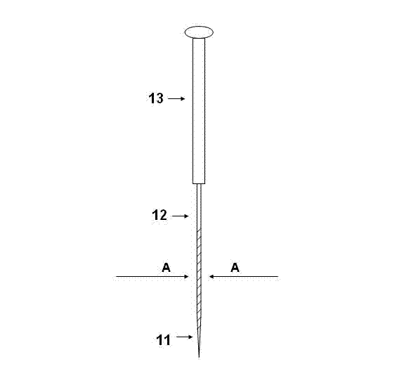
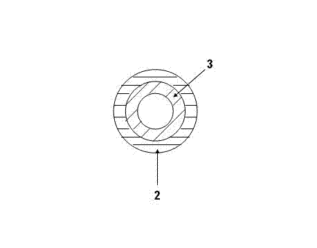
[0028] A detection microneedle with enhanced Raman and fluorescence signals, such as figure 1 As shown, the detection microneedle includes: a needle handle 13 , a needle body 12 and a needle tip 11 connected in sequence. Such as image 3 As shown, the metal nanomaterial layer 2 is covered on the surface of the needle body 12 and the needle tip 11 , and the polymer material layer 3 is coated on the metal nanomaterial layer 2 . The particle size of the metal nanomaterial is 20-1000 nm, see Figure 5 , the surface of the needle body and the needle tip is covered with 1-2 layers of gold nanoparticles, and the diameter of the gold nanoparticles is 180nm. In this example,

[0029] The polymer material layer is one of polystyrene layer, polylactic acid layer, polyurethane layer or polyethylene polypropylene fiber layer.

[0030] The metal nanomaterial layer can be one of the following:

[0031] 1) The metal nanomaterial layer is a gold nanoparticle layer, a silver nanoparticle ...

Embodiment 2

[0036] A method for preparing a detection microneedle with enhanced Raman and fluorescence signals, the steps are as follows:

[0037] Step 1) Modifying at least one of amino groups, aldehyde groups, carboxyl groups and hydroxyl groups on the surface of the needle body and needle tip of the microneedle;

[0038] Step 2) Insert the needle body and tip of step 1 into the concentration of 10 8 -10 18 After standing for 12-48 hours in the metal nanoparticle suspension of particles / liter, take out the microneedles;

[0039] Step 3) Insert the needle body and needle tip treated in step 2 into the polymer solution in which the polymer mass to solution volume ratio is 0.1-10% and let it stand for 1-60 seconds, then take it out to obtain the detection microneedle .

Embodiment 3

[0040] Example 3 Preparation of a detection microneedle with enhanced Raman and fluorescence signals

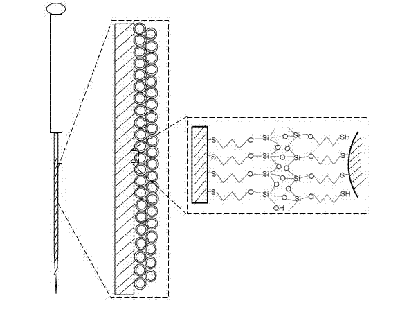
[0041] To obtain functionalized microneedles (methods for modifying thiol groups are known in the art) by modifying the surface of the needle body and tip of the detection microneedles with sulfhydryl groups, adjust the gold nanoparticle suspension to a concentration of 1×10 8-18 pcs / liter. Then the functionalized microneedle is taken out after standing in the gold nanoparticle suspension for 12-48 hours. Due to the chemical bond between the sulfhydryl group on the surface of the microneedles and the gold nanoparticles, the gold nanoparticles will be adsorbed on the surface of acupuncture needles, such as Figure 5 As shown, a nanostructure with uniform 1-2 layer structure and enhanced Raman and fluorescence signals is formed. see figure 2, the metal nanoparticle layer can be connected with the surface of the needle body and the needle tip through groups such as mercapto ...


PUM

PropertyMeasurementUnit
Particle sizeaaaaaaaaaa
Diameteraaaaaaaaaa

Abstract

The invention discloses a detecting micro-needle with a strengthened Raman and a fluorescence signal and a preparation method of the detecting micro-needle. The detecting micro-needle is characterized in that a medical acupuncture needle is used a basis for preparation. A metal nanometer material layer and a high molecular material layer with a strengthened Raman and a fluorescence signal cover the surfaces of the needle body and the needle tip part of the micro-needle. The detecting micro-needle structure comprises a micro-needle with a mercapto and amination surface, a metal nanoparticle layer and a high molecular material layer. The metal nanoparticles with a diameter of 20-1000nm coat the surface of the acupuncture needle by a covalent bond or a static adsorption effect. The high molecular layer covers the protecting metal nanoparticles in the outmost layer. Raman and fluorescence detection of in vitro samples and sampling and minimally invasive sampling and Raman and fluorescence detection in organism can be carried out based on the features of the detecting micro-needle. A novel rapid and super-sensitive detecting method for in vivo and in vitro experiment research, clinical diagnosis and large sample screening is provided by the invention.

Description

technical field [0001] The invention relates to the technical field of a functional detection microneedle, more specifically, a detection microneedle with enhanced Raman and fluorescence signals and a preparation method thereof. Background technique [0002] Monitoring the distribution of endogenous or exogenous molecules in target organs is more meaningful than measuring their concentration in blood, including the fields of understanding physiological mechanisms such as neural signal transduction and evaluating drug bioavailability such as cancer chemotherapy. Traditional in situ analysis methods cannot fully meet this monitoring requirement due to their invasiveness or complicated sample preparation. Raman spectroscopy can provide chemical and physical information based on molecular vibrations, because Raman detection does not need to consider the physical state of the sample, such as state, temperature, shape and size, so this technology is an ideal technology for real-ti...

Claims


Application Information

Patent Timeline
IPC IPC(8): A61B5/00G01N21/65G01N21/64A61K49/00
Inventor 董健陶琴许蓓蓓唐栋梁郭明德袁倩倩
Owner SOUTHEAST UNIV
Why Eureka
Try Eureka