Brain delivery method for nano-medicament carrier
A nano-drug carrier and nano-drug technology, applied in the field of medicine, can solve problems such as the inability to further realize molecular targeting of lesions in the brain, unsatisfactory efficiency, and lack of biological targeting characteristics, so as to overcome adverse in vivo properties and realize The effect of lesion targeting and enhancing the signal intensity of diagnostic molecules
- Summary
- Abstract
- Description
- Claims
- Application Information
AI Technical Summary
Problems solved by technology
Method used
Image
Examples
Embodiment 1
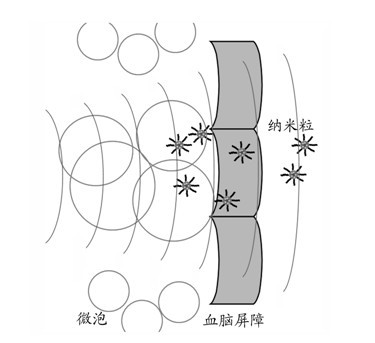
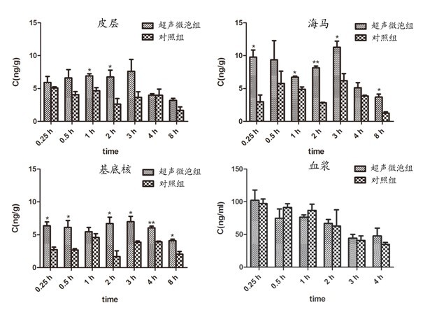
[0039] Example 1. Intracerebral delivery of coumarin-loaded 6 nanoparticles mediated by ultrasound combined with microbubbles
[0040] Preparation of coumarin-6 nanoparticles: 10-25 mg monomethoxy polyethylene glycol-polylactic acid (MPEG-PLA) block copolymer was dissolved in 1 ml dichloromethane solution (containing 0.01-0.2 mg coumarin 6), add 2 ml of 1% sodium cholate solution, and continuously sonicate at 220 W for 30 s-2 min to obtain colostrum. Add colostrum into 8-25 ml of 0.5% sodium cholate solution, stir for 5 min, remove dichloromethane by rotary evaporation, centrifuge at 14,000 rpm at 4°C for 45 min, remove supernatant, and refill with 0.01 mol / L pH7.0 HEPES Disperse and pass through a Sepharose CL-4B gel column to remove unencapsulated coumarin 6 to obtain coumarin 6 nanoparticles.
[0041] Ultrasound devices for clinical diagnosis are used, and SonoVue (particle size about 2 μm) approved by SFDA for use in my country is used as ultrasonic microbubbles. As show...
Embodiment 2
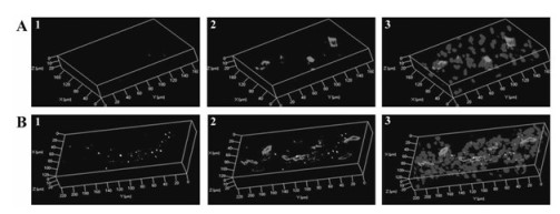
[0045] Example 2. Ultrasound combined with microbubbles mediates the transport of coumarin-6 liposomes into the brain parenchyma
[0046] Preparation of coumarin 6-loaded liposomes: 5-30 mg DMPC, 0.01-1 mg coumarin 6 were dissolved in 2 ml chloroform, and rotary evaporation was used to obtain a lipid film, and 10 ml solution was added for hydration to obtain a multilamellar liposome plastid. Small single-lamellar liposomes were prepared by ultrasonication with a 200 W probe, high-pressure homogenization or membrane extrusion, and the unencapsulated coumarin 6 was removed by gel column separation.
[0047] Using ultrasonic devices for clinical diagnosis, SonoVue is ultrasonic microbubbles. Liposomes were administered immediately after ultrasound combined with microbubble treatment. Three hours later, the experimental mice were perfused with normal saline to remove blood and fixed with 4% paraformaldehyde. The brain was taken out, fixed with 4% paraformaldehyde for 24 h, dehyd...
Embodiment 3
[0049] Example 3. Ultrasound combined with microbubbles mediates Fe 3 o 4 Cerebrovascular endothelial and neuronal cellular uptake of nanoparticles
[0050] Using ultrasonic devices for clinical diagnosis, SonoVue is ultrasonic microbubbles. Immediately after ultrasound and microbubble treatment, Fe 3 o 4 Nanoparticles, after 3 h, the heart of experimental mice was perfused with normal saline to remove the blood, and fixed with 2.0% glutaraldehyde. Take the brain, cut 1 cm from the hippocampus 3 Tissue blocks were fixed with 2% glutaraldehyde for 24 hours, fixed with osmic acid, embedded, ultra-thin sectioned, and observed by transmission electron microscopy that ultrasound combined with microbubbles mediated the opening of BBB tight junctions and the formation of transport vesicles.
[0051] The results show( Figure 4 ), there were obvious endocytic vesicles in brain vascular endothelial cells in the ultrasound combined with microbubble group, Fe 3 o 4 Nanoparticles...
PUM
| Property | Measurement | Unit |
|---|---|---|
| particle diameter | aaaaa | aaaaa |
| particle diameter | aaaaa | aaaaa |
| particle diameter | aaaaa | aaaaa |
Abstract
Description
Claims
Application Information
 - R&D
- Intellectual Property
- Life Sciences
- Materials
- Tech Scout
- Unparalleled Data Quality
- Higher Quality Content
- 60% Fewer Hallucinations
Browse by: Latest US Patents, China's latest patents, Technical Efficacy Thesaurus, Application Domain, Technology Topic, Popular Technical Reports.
© 2025 PatSnap. All rights reserved.Legal|Privacy policy|Modern Slavery Act Transparency Statement|Sitemap|About US| Contact US: help@patsnap.com


