Method for inducing human umbilical cord mesenchymal stem cells into testicular interstitial cells and application thereof

A technology of Leydig cells and Leydig stem cells is applied in inducing human umbilical cord mesenchymal stem cells to differentiate into Leydig cells and application fields, which can solve the problems of low differentiation efficiency, loss and low gonadal hormones, and achieve high differentiation efficiency, Effects of short differentiation time and strong testosterone secretion capacity

Inactive Publication Date: 2012-07-04
JINAN UNIVERSITY
View PDF1 Cites 5 Cited by
  • Summary
  • Abstract
  • Description
  • Claims
  • Application Information

AI Technical Summary

Problems solved by technology

But bone marrow mesenchymal stem cells in vitro proliferation ability, multidirectional differentiation ability and cell stability will be weakened or even lost to varying degrees with the age of the bone marrow donor or the impact of the disease; moreover, the differentiation efficiency of bone marrow mesenchymal stem cells is relatively low. Low, the amount of gonadal hormones secreted is low, and it is necessary to find other sources of stem cells and effective induction differentiation methods, which can induce the differentiation of stem cells into Leydig cells more efficiently

Method used


Image

  • Method for inducing human umbilical cord mesenchymal stem cells into testicular interstitial cells and application thereof
  • Method for inducing human umbilical cord mesenchymal stem cells into testicular interstitial cells and application thereof
  • Method for inducing human umbilical cord mesenchymal stem cells into testicular interstitial cells and application thereof

Examples


Embodiment 1

[0043] Example 1 Isolation and Expansion of Human Umbilical Cord Mesenchymal Stem Cells

[0044] Tissue block adherent separation method was used. Obtain the umbilical cord under aseptic conditions, soak the umbilical cord in 0.25% iodophor by volume for 3 minutes for disinfection, rinse with normal saline to remove blood clots on the surface of the umbilical cord and in the blood vessel, cut the umbilical cord, and tear off the Wharton’s jelly tissue around the blood vessel wall with tweezers ( Wharton's jelly), cut it into 1.5-2.5mm 3 The size of the tissue block is placed in a 24-well plate, and 800ml of culture medium is added (the culture medium contains 10% by volume of fetal bovine serum, basic fibroblast growth factor of 5ng / ml, 2mM L-glutamine, 216μg / ml NaHCO 3 , 100U / ml penicillin, 100μg / ml streptomycin and 1μg / ml amphotericin B LG-DMEM culture fluid), put in the incubator, 37 ℃, 5% CO 2 culture under static conditions. After 12-15 days, microscopic examination ...

Embodiment 2

[0045] Example 2 Cell Morphology Observation

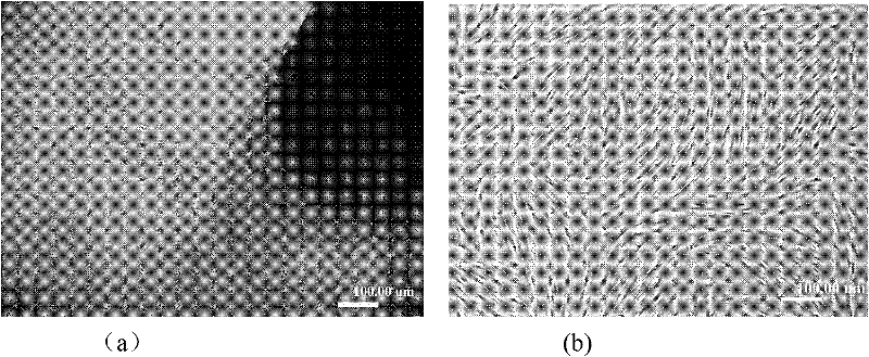
[0046] After the umbilical cord Walton jelly tissue block was inoculated into a 24-well plate and cultured for 10 days, a small amount of cells could be gradually dissociated from the tissue block, adhered to the wall, and continued to proliferate ( figure 1 a). Observed under a microscope, it can be seen that the cells are in the shape of a uniform long spindle, which is a typical shape of fibroblasts, growing in a parallel arrangement or in a spiral shape, and fused into a single layer of adherent cells with the continuous expansion of the colony growth. Conventional subculture, the cells can be seen to appear swirl after overgrowth ( figure 1 b).

Embodiment 3

[0047] Example 3 Identification of Cell Surface Antigens by Flow Cytometry

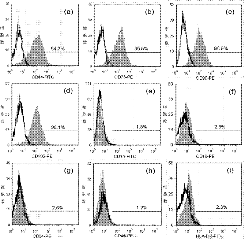
[0048] The normal cultured umbilical cord mesenchymal stem cells were taken, and flow cytometry was used to detect the expression of cell surface antigens, including CD105, CD73, CD90, CD45, CD34, CD14, CD19 and HLA-DR.

[0049] (1) After the cells grow to 80-90% confluent, they are digested with 0.25% trypsin solution with mass volume ratio, collected by centrifugation, washed twice with PBS solution, and the concentration of the cell suspension is adjusted to 10 6 cells / ml.

[0050] (2) Add 2 ml of PBS solution containing 2.5% FBS by volume, mix the cells by pipetting, and centrifuge at 200 g.

[0051] (3) Antibodies used include CD105, CD73, CD90, CD45, CD34, CD14, CD19 and HLA-DR antibodies. Non-specific backgrounds were incubated with mouse anti IgG1 / PE and mouse anti IgG1 / FITC as a control. The antibody was diluted with PBS solution at a ratio of 1:50 by volume, and incubated on ice in the dar...


PUM


Abstract

The invention discloses a method for inducing human umbilical cord mesenchymal stem cells into testicular interstitial cells and application thereof. The method comprises the following steps: cloning mouse steroidogenic factor-1 (SF-1) gene into an adenovirus shuttle plasmid pAdtrack-CMV-EGFP to construct a recombinant vector pAdtrack-CMV-SF-1; recombining the recombinant vector pAdtrack-CMV-SF-1 and an adenovirus carrier plasmid pAdEasy-1 to obtain an adenovirus plasmid pAd-SF-1 carrying SF-1 gene; digesting and linearizing the adenovirus plasmid pAd-SF-1, and packaging with human embryonic kidney cells AD-293 to obtain adenovirus; adding the adenovirus into an induction culture medium to infect human umbilical cord mesenchymal stem cells, and carrying induced differentiation of the human umbilical cord mesenchymal stem cells to testicular interstitial cells. Through the induction using the method of the invention, human umbilical cord mesenchymal stem cells can in vitro differentiate into testicular interstitial cells only in one week, so as to provide an important cell source for treatment of testosterone deficiency using cell replacement or genetic method.

Description

technical field [0001] The invention belongs to the field of biotechnology of inducing stem cell differentiation, and in particular relates to a method and application for inducing human umbilical cord mesenchymal stem cells to differentiate into testicular Leydig cells. Background technique [0002] With the prolongation of life expectancy and the problem of population aging, the number of middle-aged and elderly male partial androgen deficiency (PADAM) patients is increasing. At present, exogenous androgen replacement therapy (ART) is mainly used clinically, the purpose is to maintain the physiological concentration of testosterone in serum to replace the physiological function of endogenous testosterone. However, long-term use of androgen has relatively large side effects. Firstly, there are many target cells of androgen in the body. Long-term use of exogenous androgen therapy is prone to toxic and side effects, especially in the prostate gland; Patients need frequent bl...

Claims


Application Information

Patent Timeline
Patent Type & Authority Applications(China)
IPC IPC(8): C12N5/10C12N15/12C12N15/861C12N7/01C12N5/0775C07J1/00
Inventor 魏星彭公峰
Owner JINAN UNIVERSITY
Why Eureka
Try Eureka