
Tissue engineering cartilage framework material, as well as preparation method and device thereof

A scaffold material and tissue engineering technology, used in bone implants, medical science, prostheses, etc., can solve the problem of not having the characteristics of the calcification layer of the fibrous structure arranged horizontally on the surface of the cartilage, affecting the fusion of the graft and the surrounding tissue, increasing the amount of cartilage compressive strength and other issues, to achieve the effect of improving wear resistance, preventing moisture loss, and maintaining high elastic modulus

Active Publication Date: 2012-07-18
西安博鸿生物技术有限公司
View PDF15 Cites 9 Cited by
  • Summary
  • Abstract
  • Description
  • Claims
  • Application Information

AI Technical Summary

Problems solved by technology

Although Chinese patent application 200910117158.6 has a layered structure, its structure is only different in the content of hydroxyapatite, and the cartilage scaffold material does not have a vertically oriented structure that can increase the compressive strength of cartilage
The cartilage repair material constructed in Chinese patent application 201010297976.1 is in the longitudinal arrangement of natural cartilage matrix, but it does not have the horizontal arrangement of fibrous structure on the surface of cartilage and the composition characteristics of the calcified layer
Chinese patent application 200510012257.X uses materials such as polyvinyl alcohol (PVA) and hydroxyapatite to construct multilayer cartilage materials, simulating the gradual change of natural cartilage components in the multilayer structure, and replacing cartilage tissue with polymer materials such as PVA. collagen, proteoglycan, etc., but the degradation and metabolism of polymer materials in the body is completely different from that of cartilage matrix. The degradation of the former will produce acidic products, causing non-bacterial inflammatory reactions, and directly affecting the fusion of grafts and surrounding tissues.

Method used

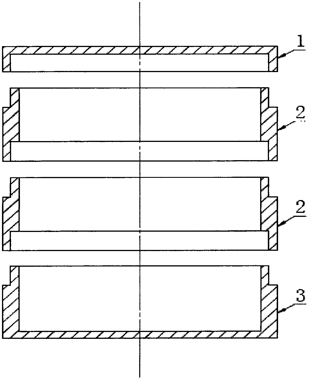

Image

  • Tissue engineering cartilage framework material, as well as preparation method and device thereof
  • Tissue engineering cartilage framework material, as well as preparation method and device thereof
  • Tissue engineering cartilage framework material, as well as preparation method and device thereof

Examples


Embodiment 1

[0027] Step 1. Preparation of natural cartilage matrix: 10-200mm 3 Mammalian articular cartilage pieces of the same size without perichondrium are frozen in liquid nitrogen for 3 hours, then transferred to a pre-cooled pulverizer for pulverization, dissolved in acetic acid solution with a volume concentration of 1%, and vacuum freeze-dried to obtain cartilage matrix;

[0028] Step 2, preparation of calcified layer: dissolve freeze-dried cartilage matrix, GAG and X-type collagen in 1% acetic acid solution so that the final concentrations are 5 mg / ml, 8 mg / ml and 5 mg / ml respectively, and use hydrogen Adjust the pH of the calcium oxide solution to neutral to obtain a cartilage matrix solution; fully mix the calcined bone powder (0.2 mm in particle size) with the cartilage matrix solution at a concentration of 0.4 g / ml, inject it into the bottom box of the culture vessel, and make the thickness of the gel layer 4 mm, add Cover and wait for the gel solution to solidify at 37°C, t...

Embodiment 2

[0033] Step 1. Preparation of natural cartilage matrix: 10-200mm 3 Mammalian articular cartilage pieces of the same size without perichondrium are frozen in liquid nitrogen for 3 hours, then transferred to a pre-cooled pulverizer for pulverization, dissolved in acetic acid solution with a volume concentration of 1%, and vacuum freeze-dried to obtain cartilage matrix;

[0034] Step 2, preparation of calcified layer: dissolve freeze-dried cartilage matrix, GAG and X-type collagen in 1% acetic acid solution so that the final concentrations are 10 mg / ml, 9 mg / ml and 10 mg / ml respectively, and use hydrogen Adjust the pH of the calcium oxide solution to neutral to obtain the cartilage matrix solution; fully mix the calcined bone powder (Bayameng brand hydroxyapatite bone powder particle size 0.1mm) and the cartilage matrix solution at a concentration of 0.15g / ml, and pour it into the bottom of the culture vessel box, so that the thickness of the gel layer is 2mm, and after the gel ...


PUM

PropertyMeasurementUnit
Thicknessaaaaaaaaaa
The inside diameter ofaaaaaaaaaa
Depthaaaaaaaaaa

Abstract

The invention discloses a tissue engineering cartilage framework material. The cartilage framework material provided by the invention sequentially comprises a surface layer, a middle layer and a calcification layer from the top down; the surface layer is composed of freeze-dry cartilage matrix and II-type collagen, and matrix fiber is in parallel distribution in the horizontal direction; the middle layer is composed of freeze-dry cartilage matrix, GAG, X-type collagen, II-type collagen and TGF-Beta, and matrix fiber is arranged longitudinally; and the calcification layer is composed of freeze-dry cartilage matrix, GAG, X-type collagen and sintered bovine bone power, and fiber is distributed in a three dimensional staggered manner. The prepared cartilage framework material has a cartilage repairing effect, is favorable to the integration of the framework material and subchondral bone after the framework material is planted, and promotes the regeneration of the subchondral bone; after cartilage cell compound, the framework material can be used for full-layer cartilage defect repairing, the regenerated cartilage tissue has the space structure and protein components, which are the same with those of natural cartilage; and the framework material is suitable for repairing cartilages in load bearing joints such as knee joints and the like, cartilage with higher regenerated compressive strength can be regenerated, and the abrasion performance and anti-pressure capability of the surface of the cartilage framework are enhanced.

Description

technical field [0001] The invention belongs to the technical field of tissue engineering medical biomaterials, and in particular relates to a tissue engineering cartilage scaffold material, a preparation method and a preparation device thereof. Background technique [0002] Articular cartilage injury is a common clinical disease. Domestic and foreign surveys show that the prevalence of articular cartilage damage is about 15%, the prevalence rate of people aged 40 is 10% to 17%, and that of people over 60 years old is 50%, while 80% of people over 75 years old suffer from skeletal joints. Cartilage defects associated with inflammation. In the United States, articular cartilage damage is the second leading cause of disability in men over the age of 50 (after ischemic heart disease) and the leading cause of chronic disability in those older than middle age. According to data from the American College of Rheumatology (ACR), there are currently about 20 million patients with a...

Claims


Application Information

Patent Timeline
IPC IPC(8): A61L27/40A61L27/36A61L27/24A61L27/20A61F2/28
Inventor 姚瑶
Owner 西安博鸿生物技术有限公司
Learn More