Chordoma cell line and application thereof
A technology for chordoma and its application, which is applied in the field of establishment of human chordoma cell lines, and can solve problems such as unreported
- Summary
- Abstract
- Description
- Claims
- Application Information
AI Technical Summary
Problems solved by technology
Method used
Image
Examples
Embodiment 1
[0036] Embodiment 1: the establishment of human chordoma cell line
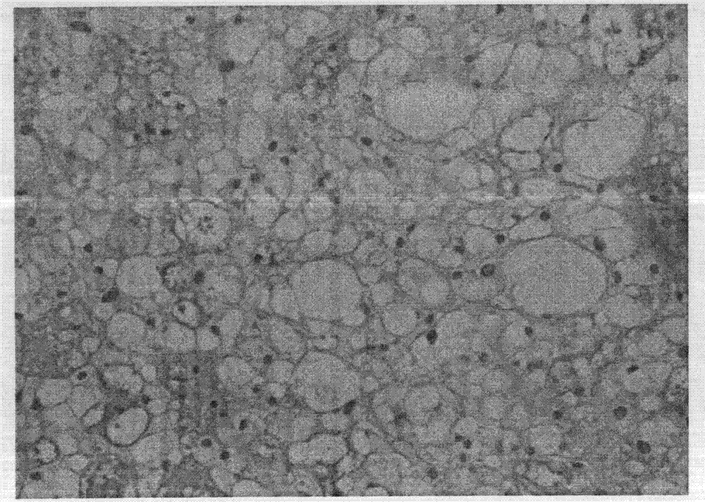
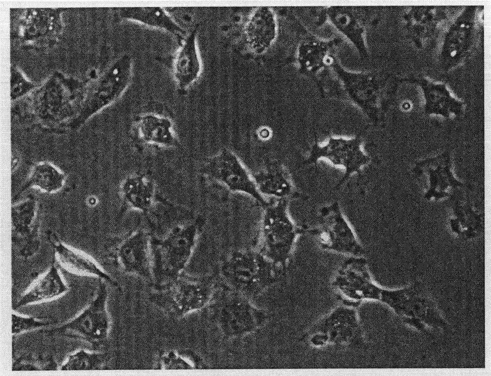
[0037] Primary culture of HBC2: the cell line in the present invention is obtained by separating and culturing the skull base chordoma tissue of the surgical patient. The tumor tissue was obtained from a surgical specimen of a patient in the Neurosurgery Center of Beijing Tiantan Hospital Affiliated to Capital Medical University. The pathological diagnosis was brain chordoma. Under the microscope, the cells were arranged in sheets, with round or oval nuclei. Soap-like structure ( figure 1 ). The specific method is as follows: wash the tissue block twice with Hank’s (1:1) containing red solution in a sterile ultra-clean bench, remove blood vessels and necrotic tissue, and cut the tissue block into about 1 mm with tissue scissors 3 Size, add 3ml 0.04% EDTA / 0.5% trypsin, digest in a 37°C water bath for 10 minutes, observe under a microscope, after digesting into single cells, add 300ul fetal bovine serum to st...
Embodiment 2
[0041] Example 2: Observation of Ultrastructure of Cells by Transmission Electron Microscopy
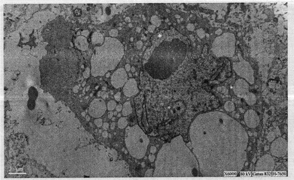
[0042] When the HBC2 cells were subcultured for 14 generations, when the cells reached 90% confluence, discard the culture medium, add 3ml 0.1M PBS (PH7.4), scrape the cells with a rubber cell scraper, collect the cells, centrifuge (800rpm / min, 10 Minutes), discard the supernatant, pre-fix with glutaraldehyde / osmium tetroxide for 2 hours, wash 3 times with 0.1M PBS (pH7.4), 10 minutes each time. Then the specimens were post-fixed, dehydrated, soaked, embedded, cut into ultra-thin sections and stained with heavy metals (completed by the electron microscope room of Beijing Institute of Neurosurgery).
[0043] The ultrastructure of HBC2 was observed by transmission electron microscopy. It can be seen that there are a large number of microvilli on the surface of the cell membrane, the shape of the nucleus is irregular, the nuclear membrane is sunken, the nucleolus is large and obvious, a...
Embodiment 3
[0044] Example 3: Detection of marker proteins derived from notochord tissue by immunohistochemical staining
[0045] When the HBC2 cells were subcultured for 14 generations, after the cells reached 90% confluence, they were washed twice with Hank's. Add 3 ml of 0.04% EDTA / 0.5% trypsin to digest for about 2 minutes, add 2 ml of serum-containing medium to terminate the reaction, centrifuge (800 rpm / min, 10 minutes) and wash once, and collect the precipitated cells at the bottom of the centrifuge tube. Resuspend the cells in 5ml of DMEM / F12 culture medium containing 20% serum, inoculate them on a culture dish covered with coverslips, and discard the culture medium after the cells grow to 90% confluence, and use 0.1M PBS (PH7.4) Wash 3 times, add cold acetone to fix at 4°C for 10 minutes, wash 3 times with 0.1M PBS (PH7.4), 5 minutes each time, 3% hydrogen peroxide for 10 minutes at room temperature (to eliminate endogenous peroxidase). Wash 3 times with 0.1MPBS (PH7.4), 5 min...
PUM
 Abstract
Description
Claims
Application Information
 - R&D
- Intellectual Property
- Life Sciences
- Materials
- Tech Scout
- Unparalleled Data Quality
- Higher Quality Content
- 60% Fewer Hallucinations
Browse by: Latest US Patents, China's latest patents, Technical Efficacy Thesaurus, Application Domain, Technology Topic, Popular Technical Reports.
© 2025 PatSnap. All rights reserved.Legal|Privacy policy|Modern Slavery Act Transparency Statement|Sitemap|About US| Contact US: help@patsnap.com


