Method for preparing complex organ precursor with branch vessel network

A branch and network technology, applied in prosthetics, medical science, etc., can solve the problems of no-channel differentiation of blood vessels, uneven distribution, low similarity, etc., achieve excellent biocompatibility, overcome the surface and internal structure of three-dimensional stents The effect of simplicity and excellent mechanical properties

Inactive Publication Date: 2012-08-15
TSINGHUA UNIV
View PDF3 Cites 17 Cited by
  • Summary
  • Abstract
  • Description
  • Claims
  • Application Information

AI Technical Summary

Problems solved by technology

[0006] The purpose of the present invention is to provide a method for preparing complex organ precursors with branched vascular networks, aiming to achieve accurate positioning of various cells and scaffold materials in space by using a combined mold for step-by-step perfusion on the basis of previous work. Forming a composite tissue and organ precursor of multiple cells and multiple growth factors wrapped in a multi-layer polymer material shell; using the principles of mold combination and polymer coagulation forming to realize the reconstruction of complex tissues and organs, the invention can form The complex three-dimensional structure containing different cell matrix materials, branched vascular channels and multi-layer synthetic polymer shell overcomes the simple surface and internal structure of the three-dimensional scaffold in tissue engineering, low similarity to real organs, single cell types and uneven distribution, Closed cells are not easy to survive, it is difficult to differentiate blood vessels without channels, and the shape of the stent is limited, etc.

Method used


Image

  • Method for preparing complex organ precursor with branch vessel network
  • Method for preparing complex organ precursor with branch vessel network
  • Method for preparing complex organ precursor with branch vessel network

Examples


Embodiment 1

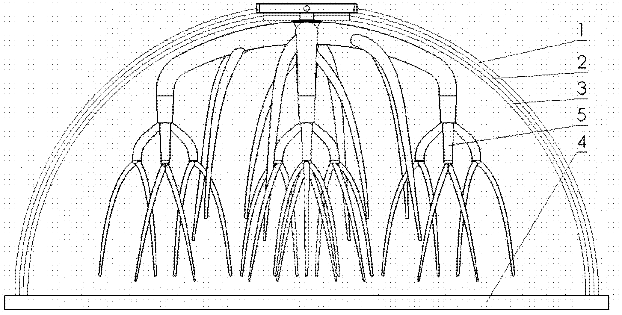
[0041] Embodiment 1: 1) adopt stainless steel material to prepare three-layer shell mold and bottom mold, and described shell mold surface is spherical, adopts polytetrafluoroethylene material to prepare double-layer branch mold with rapid prototyping method; 2) prepare fibrinogen solution, Inject a small amount of mixture of gelatin / fibrinogen and endothelial cells into the combined mold with a cell density of 1×10 7 1 / mL, inject thrombin solution (20IU / mL) to make the cell / natural polymer material layer form the bottom stable structure; 3) inject the mixture of gelatin / fibrinogen, endothelial cells and smooth muscle cells into the composite mold, and the cell density is 1×10 7 cells / mL, adding hepatocyte growth factor (HGF0.5ng / mL), human platelet-derived growth factor (BB or PDGF-BB 50ng / mL), transforming growth factor β1 (TGFβ1 10ng / mL) and basic fibroblast growth Factor (b-FGF 2.5ng / mL). Make the cell natural polymer material evenly distributed, inject thrombin solution...

Embodiment 2

[0042] Embodiment 2: 1) adopt silicon rubber material to prepare double-layer shell mold and bottom mold, described shell mold surface is ellipsoid, adopt PLGA material to prepare double-layer branch mold with rapid prototyping method; 2) prepare fibrinogen solution, in Inject a small amount of mixture of gelatin / fibrinogen and adipose-derived stem cells into the combination mold, and the cell density is 1×10 7 cells / mL, add endothelial cell growth factor and inject thrombin solution (20IU / mL) to make the cell / natural polymer material layer form the bottom stable structure; 3) prepare fibrinogen / endothelial cell mixture containing 1% paclitaxel, cell density 1×10 6 pcs / mL, poured into the combination mold, inject thrombin solution (20IU / mL) to make the cell / natural polymer material layer form an upper stable structure; 4) remove the shell mold and branch mold together, and peel off layer, recombine the molds, inject the mixture of gelatin / fibrinogen and adipose-derived stem c...

Embodiment 3

[0043] Embodiment 3: 1) adopt polytetrafluoroethylene material to prepare three-layer shell mold and bottom mold, described shell mold is the irregular shape of similar organ surface, adopts polyurethane material to prepare branch mold with rapid prototyping method; 2) preparation 1% Collagen / endothelial cell mixture in sodium citrate (cell density 1 x 10 7 cells / mL), poured into the combination mold, and placed at 37°C for 10 minutes to stabilize the structure of the collagen / endothelial cell mixture; 3) Remove the shell mold and the branch mold together, and take the first shell mold and the second shell mold. Buckle the shell mold back onto the bottom mold, prepare a polylactic acid / isopropanol solution with a concentration of 30%, add 30% sodium citrate, stir evenly, pour it into the peripheral gap left by the third shell mold, Use the PBS extraction method to form the inner layer of synthetic polymer material shell; 4) buckle the first layer of shell mold back on the bott...


PUM


Abstract

The invention provides a method for preparing complex organ precursor with a branch vessel network. The method comprises the following steps: firstly preparing one or more cellular matrix solutions and a synthetic macromolecular solution; pouring the cellular matrix solution into a combined mold in layers for physical or chemical cross-linking to form a multilayered cellular matrix layer; taking out a branch mold and removing a shell mold in layers from inwards to outwards; pouring macromolecular solutions of different types into gaps remained in the shell mold respectively so as to form a multilayered synthetic macromolecule shell; and removing the combined mold and abutting two formed structures so as to obtain the complex organ precursor. According to the method, a complex organ precursor three-dimensional structure, in which a branch vessel network is provided and cellular matrix materials of different types and synthetic macromolecule support shells are combined, is formed; and the defects that the tissue engineering is simple in three-dimensional support surface and the internal structure, low in similarity with real organs, single in cell type and uneven in cell distribution, less in possibility for survival of closed cells, difficult in non-channel differentiation vessels and strong in support appearance limitation are solved.

Description

technical field [0001] The invention belongs to the technical field of artificial manufacturing of biological tissues and organs, in particular to a process for preparing tissue and organ precursors by using synthetic polymer materials and cell matrix materials, and belongs to the technical field of biological tissue engineering. Background technique [0002] Tens of millions of patients suffer from tissue defect or organ failure every year in the world. However, living donor organs are limited, and existing mechanical devices do not have all the functions of the organs and cannot prevent further deterioration of the patient's condition. Accordingly, the tissue engineering (Tissue Engineering) technology for the purpose of improving the treatment level of such diseases came into being. [0003] Tissue engineering was officially proposed and determined by the National Science Foundation of the United States in 1987. It is the application of the principles of cell biology, bi...

Claims


Application Information

Patent Timeline
Patent Type & Authority Applications(China)
IPC IPC(8): A61L27/38A61L27/26
Inventor 王小红
Owner TSINGHUA UNIV
Why Eureka
Try Eureka