Method for proliferating influenza A viruses
A type A influenza virus and influenza virus technology, applied in the field of animal infectious diseases, can solve the problems of foreign virus contamination, antigen variation, and insufficient number of chicken embryos
- Summary
- Abstract
- Description
- Claims
- Application Information
AI Technical Summary
Problems solved by technology
Method used
Image
Examples
Embodiment 1
[0103] Example 1: Isolation and Identification of Novel Type A H1N1 Influenza Virus
[0104] collection of samples
[0105]Tracheal cotton swabs of pigs suspected of swine influenza infection in a pig farm in Jiangxi Province, China were collected, and the swabs were placed in sterile saline containing 100 U / mL penicillin and streptomycin. The collected samples were immediately stored at 2-8°C, and sent for inspection within 24 hours, and stored at -70°C for long-term storage.
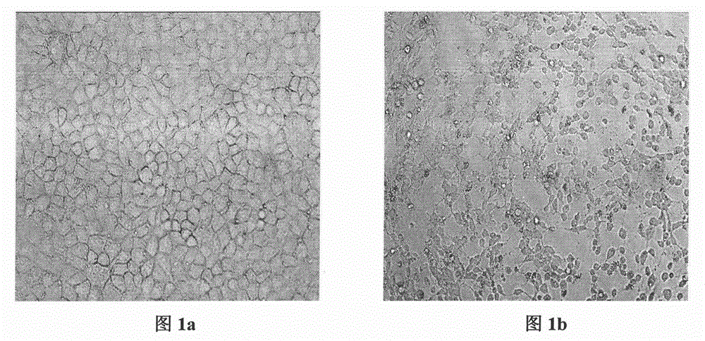
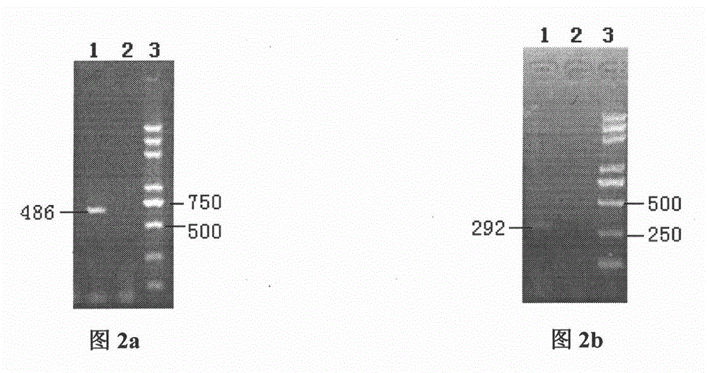
[0106] Virus isolation and identification
[0107] For the preparation of 80% sheets of cells, observe the growth state of the cells with a 40x microscope, select the MDCK cells in the logarithmic growth phase, pour out the cell growth solution gently, and wash the cells twice with sterilized saline to remove the cell surface newborn bovine serum. Add the cell maintenance solution containing 2 μg / mL TPCK-trypsin.
[0108] For the inoculation of cell culture wells, MDCK cells were used to isolate th...
Embodiment 2
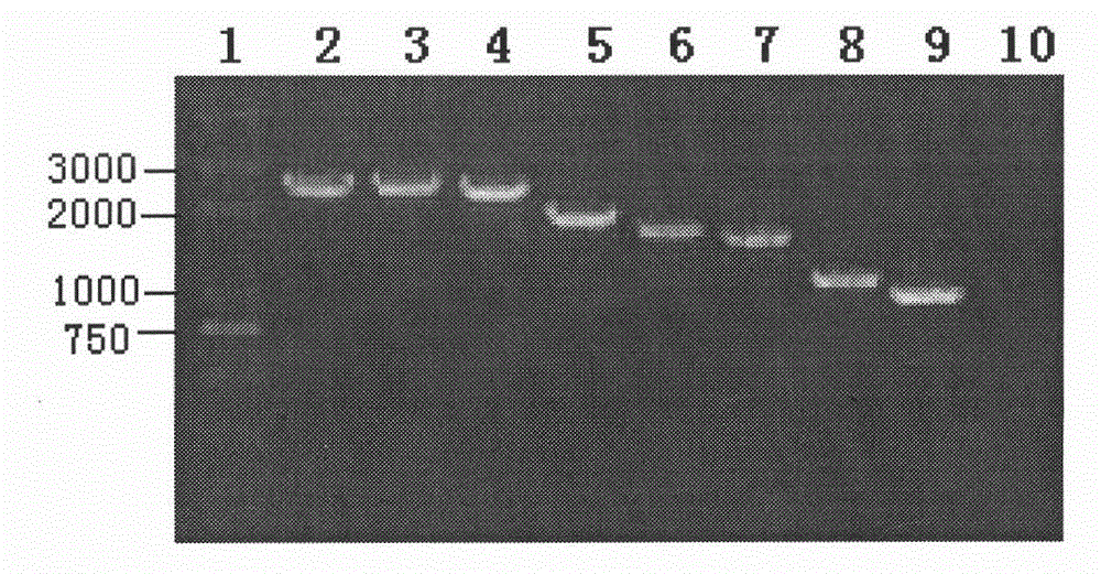
[0154] Example 2: Whole Genome Sequencing of New Type A H1N1 Influenza Virus
[0155] Sequencing the whole genome of influenza A H1 virus, the steps are as follows:
[0156] Using the cDNA described in step 2) as a template, amplify the full length of 8 gene sequences of influenza A H1 virus, i.e. HA, M, NA, NP, NS, PA, PB1 and PB2, and the amplification obtained as sequence table SEQ The nucleotide sequence shown in ID NO: 1-8 (its sequence is such as (Genbank accession number is corresponding to JF275925-JF275932)), the nucleotide sequence of the specific primer that amplifies above-mentioned 8 genes is as follows:
[0157] Amplify the HA gene:
[0158] P15'AGCAAAAGCAGGGG 3',
[0159] P25'AGTAGAAACAAGGGTGTTTT3';
[0160] Amplify the M gene:
[0161] P15'AGCAAAAGCAGGTAG 3',
[0162] P25'AGTAGAAACAAGGTAGTTTTTT 3';
[0163] Amplify the NA gene:
[0164] P15'AGCAAAAGCAGGAGT 3'
[0165] P25'AGTAGAAACAAGGAGTTTTTT3';
[0166] Amplify the NP gene:
[0167] P15'AGCAAAAGCAGG...
Embodiment example 3
[0188] Implementation Case 3 Effects of Different Infection Doses on Proliferation of Influenza A H1 Subtype Influenza Virus in MDCK Cells
[0189] 1) In a 12-well plate, carry out the proliferation test of influenza A H1 subtype virus of different infection doses in MDCK cells, get the 12-well plate (diameter of each well of 2.2 cm, surface area of 3.8 cm) covered with MDCK cells 2 ), the cell density is about 2×10 5 piece / cm 2 , discard the medium, and wash 3 times with PBS buffer to remove residual medium. The concentration of H1 subtype influenza virus particles is about 1×10 7 PFU / mL. MDCK cells were inoculated respectively according to the virus multiplication of infection (Multiplication of Infection, MOI) MOI=(0.3, 0.03, 0.003), which is equivalent to HA titer of 8log 2 The virus stock solution was diluted to 10 -1 , 10 -2 , 10 -3 250 μL per well, each dose connected to 3 wells, in CO 2 Adsorbed in the incubator at 37°C for 1.5h, discarded the virus solution,...
PUM
 Abstract
Description
Claims
Application Information
 - R&D
- Intellectual Property
- Life Sciences
- Materials
- Tech Scout
- Unparalleled Data Quality
- Higher Quality Content
- 60% Fewer Hallucinations
Browse by: Latest US Patents, China's latest patents, Technical Efficacy Thesaurus, Application Domain, Technology Topic, Popular Technical Reports.
© 2025 PatSnap. All rights reserved.Legal|Privacy policy|Modern Slavery Act Transparency Statement|Sitemap|About US| Contact US: help@patsnap.com


