Fusion protein and fusion protein expression vector thereof

A fusion protein and expression vector technology, applied in the field of fusion proteins, can solve the problems of difficulty in obtaining soluble active protein, difficulty in preparation, and high cost, and achieve the effects of facilitating high-density fermentation and culture, low nutritional requirements, and high-efficiency expression

Active Publication Date: 2012-12-12
THE SECOND AFFILIATED HOSPITAL ARMY MEDICAL UNIV
View PDF2 Cites 6 Cited by
  • Summary
  • Abstract
  • Description
  • Claims
  • Application Information

AI Technical Summary

Problems solved by technology

The current related researches are firstly using Escherichia coli prokaryotic expression system, which can obtain high-yield expressed protein, but its protein expression form is mostly inclusion body, and it is difficult to obtain a large amount of soluble active protein; secondly, using cell eu

Method used


Image

  • Fusion protein and fusion protein expression vector thereof
  • Fusion protein and fusion protein expression vector thereof
  • Fusion protein and fusion protein expression vector thereof

Examples


Embodiment 1

[0099] Example 1 : Synthesis of genes and primers

[0100] 1. Synthesis of 1exCAR and IgG1 Fc gene

[0101] According to the human-derived CAR extracellular region gene coding sequence (exCAR) (AccessionNumber: NM_001338) and IgG1 Fc segment gene coding sequence (Accession Number: AF237583) in GenBank, exCAR and IgG1 Fc segment were separated according to the partial tropism of Pichia pastoris genetic code The gene sequence was codon-optimized, and Shanghai Sangon synthesized exCAR and IgG1 Fc genes respectively and constructed the engineering bacteria PUC-exCAR / DH 5α , PUC-Fc / DH 5α , the nucleic acid sequence after codon optimization is as follows:

[0102] exCAR (651bp): SEQ ID NO: 1

[0103] ttgtccatcactactccagaagatgattgagaaggctaagggtgagactgcctacttgccatgtaagttcactttgtctccagaagaccaaggtccattggacatcga

[0104] gtggttgatttccccagctgacaatcagaaggttgatcaagtcattattttgtactctggtgacaagatttacgacgactactacccagacttgaagggtagaggttcacttc

[0105] acctccaatgacttgaagtctggtgatgcttctatcaat...

Embodiment 2

[0148] Example 2 : Recombinant engineered bacteria pPIC3.5K-exCAR / DH 5α construction and identification of

[0149] 2.1 exCAR gene EcoR I / SnaB I digestion

[0150] The PUC-exCAR / DH constructed in Example 1.2 5α Bacteria were inoculated in LB (AMP r ) medium, cultivate overnight at 37°C and 180 rpm, extract the PUC-exCAR plasmid with a plasmid extraction kit, and perform step-by-step enzyme digestion with EcoR I and SnaB I.

[0151] ①EcoR I digestion reaction system is as follows:

[0152]

[0153]

[0154] Place at 37°C, and after 3 hours of reaction, directly add SnaB I and corresponding buffer to the system for the second step of digestion.

[0155] ②SnaB I digestion reaction system is as follows:

[0156]

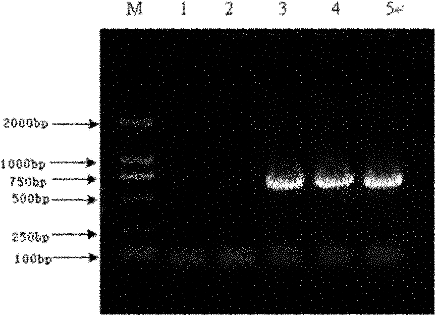
[0157] Set at 37°C and react for 3 hours. The digested product was electrophoresed in 1% agarose gel to recover the exCAR digested gene fragment.

[0158] 2.2 pPIC3.5K plasmid EcoR I / SnaB I digestion

[0159] The plasmid extraction kit extracts the pPIC...

Embodiment 3

[0171] Example 3 : Recombinant engineering bacteria pPIC3.5K-exCAR:Fc / DH 5α build

[0172] 3.1 pPIC3.5K-exCAR plasmid EcoR / Not I double digestion

[0173] The recombinant engineered bacteria plasmid pPIC3.5K-exCAR identified as positive by PCR in Example 2.6 was extracted using a plasmid extraction kit, and subjected to EcoR I / Not I double digestion.

[0174] The enzyme digestion system is as follows:

[0175] Place at 37°C and react for 3 hours. The digested product was electrophoresed in 1% agarose gel, and the digested plasmid pPIC3.5K-exCAR was recovered.

[0176] 3.2 Fc gene Not I / EcoR I double digestion

[0177] The PUC-Fc / DH constructed in Example 1.2 5α Bacteria were inoculated in LB (AMP r ) medium, cultivate overnight at 37°C and 180rpm, extract the PUC-Fc plasmid with a plasmid extraction kit, and perform Not I / EcoR I double enzyme digestion. The reaction system is as follows:

[0178]

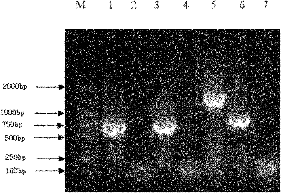
[0179] Place at 37°C and react for 3 hours. The digested product w...


PUM

PropertyMeasurementUnit
Molecular weightaaaaaaaaaa

Abstract

The invention provides a fusion protein and a fusion protein expression vector thereof. The fusion protein comprises human-derived Coxsackie virus-adenovirus receptor extracellular region and Fc fragment of human IgG1. The gene sequence of the fusion protein is subjected to codon optimization. The fusion protein expression vector comprises an optimized gene sequence of the fusion protein, and a pPIC3.5K plasmid gene sequence. The fusion protein can be combined with high affinity with Coxsackie virus/adenovirus, and can prevent the Coxsackie virus/adenovirus from infecting body cells especially cells expressing CAR, such as myocardial cells. The fusion protein provided by the invention can be used in treatments of Coxsackie virus and/or adenovirus infectious diseases.

Description

technical field [0001] The invention relates to a fusion protein and a fusion protein expression vector, in particular to a fusion protein comprising the extracellular region of human Coxsackie virus-adenovirus receptor and the Fc segment of human IgG1. Background technique [0002] Coxsackie enterovirus, belonging to the Picornavirus family, is a positive-strand RNA enterovirus. Often cause diarrhea in children, can lead to aseptic meningitis, epidemic chest pain, myocarditis, pericarditis, hand, foot and mouth disease and other diseases, infection during pregnancy can cause non-paralytic poliomyelitis lesions, and cause fetal intrauterine infection and teratogenic. At present, it is found that Coxsackie B enterovirus (CVB) often causes viral myocarditis (VMC) in children, and is the most common pathogen of VMC in children and adolescents, accounting for about 50% of myocarditis caused by various viruses . Adenovirus (adenovirus, Ad) is a kind of non-enveloped double-str...

Claims


Application Information

Patent Timeline
IPC IPC(8): C07K19/00C12N15/62A61K38/17A61P31/14C12N15/81C12N1/19C12R1/84
Inventor 张克斌钱桂生周世文黄春基余华吕胜青盛哈蕾何晓梅熊竣智
Owner THE SECOND AFFILIATED HOSPITAL ARMY MEDICAL UNIV
Why Eureka
Try Eureka