Fluorescent immune chromatographic test strip for quantitively detecting troponin I and preparation method thereof

A fluorescent immunochromatography, troponin technology, applied in biological testing, measuring devices, analytical materials, etc., can solve problems such as expensive equipment, unsuitable for single-person and small batch detection, and inability to achieve accurate quantification. Simple operation, shorten the detection time, and improve the effect of sensitivity

Inactive Publication Date: 2012-12-26
北京乐普诊断科技股份有限公司
View PDF12 Cites 25 Cited by
  • Summary
  • Abstract
  • Description
  • Claims
  • Application Information

AI Technical Summary

Problems solved by technology

Colloidal gold immunochromatography is the most widely used clinically due to its simple operation. The disadvantage is that it can only achieve semi-quantitative and cannot achieve accurate quantification; and ELISA is widely used in hospital labora

Method used


Image

  • Fluorescent immune chromatographic test strip for quantitively detecting troponin I and preparation method thereof
  • Fluorescent immune chromatographic test strip for quantitively detecting troponin I and preparation method thereof
  • Fluorescent immune chromatographic test strip for quantitively detecting troponin I and preparation method thereof

Examples


Embodiment 1

[0033] The preparation of the fluorescent immunochromatography test strip of embodiment 1 troponin I (referring to Fig. 1)

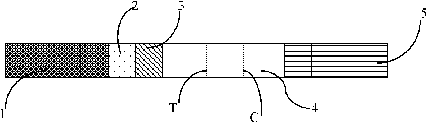
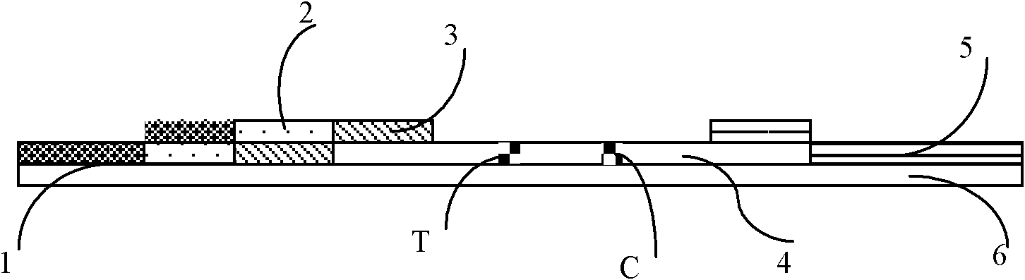
[0034]Referring to accompanying drawing 1, the fluorescent immunochromatography test strip of troponin I of the present embodiment comprises the nitrocellulose membrane 4 that is coated with quality control band (C) and detection band (T) on base plate 6, base plate, High-strength absorbent paper covering one side of the nitrocellulose membrane, 5 monoclonal antibody pads of fluorescently labeled troponin I covering the other side of the nitrocellulose membrane, 2 monoclonal antibody pads of biotin-labeled troponin I Clone antibody pad 3 and sample pad 1;

[0035] The detection zone T is coated with streptavidin; the quality control zone is coated with rabbit anti-mouse IgG antibody.

[0036] The test strip preparation method of the present embodiment comprises the following steps:

[0037] A. Antibody preparation: select commercial troponin I monoclon...

Embodiment 2

[0048] Except for the preparation step of the fluorescent microsphere mat labeled with troponin I monoclonal antibody: fluorescent microspheres with a diameter of 210 nm were selected, and other steps were the same as in Example 1.

Embodiment 3

[0050] Except that in the preparation step of the fluorescent microsphere pad labeled troponin I monoclonal antibody, the amount of 4 μl / cm was sprayed on the fluorescent microsphere pad, other steps were the same as in Example 1.


PUM

PropertyMeasurementUnit
Diameteraaaaaaaaaa
Diameteraaaaaaaaaa
Diameteraaaaaaaaaa

Abstract

The invention relates to the field of detection in clinical immunology, in particular to a fluorescent immune chromatographic test strip for quantitively detecting troponin I and a preparation method thereof. The test strip provided by the invention comprises a bottom plate, sample absorbing pads attached to the bottom plate and staggered to each other sequentially, a labeled antibody pad, a coating film and a water absorbing pad. The labeled antibody pad is a troponin monoclonal antibody pad I coated with a fluorescent micro-spherical label and a troponin monoclonal antibody pad II coated with a biotin label for identifying different epitopes. The coating film is provided with a detection strip and a quality control strip. Avidin is fixedly arranged on the detection strip. A rabbit antimouse IgG antibody is fixedly arranged on a quality control line. By improving the test strip, a biotin-avidin amplifying system and a fluorescent immune chromatographic technology are introduced into detection of troponin I. Combined with a fluorescent detector, individual quantitive detection of troponin I is realized, therefore, extreme convenience is provided for clinical use.

Description

technical field [0001] The invention relates to the field of clinical immunology detection, in particular to a fluorescent immunochromatographic test strip for quantitative detection of troponin I and a preparation method thereof. Background technique [0002] Myocardial infarction is one of the leading causes of death and disability worldwide. Rapid identification and timely treatment of patients with early acute myocardial infarction are the key to improving their survival rate. For AMI patients without typical chest pain and ECG changes, the detection of serum myocardial markers is a necessary basis for the diagnosis of AMI. The criteria for myocardial infarction defined by the World Health Organization (WHO) in the past included ischemic symptoms, abnormal changes in electrocardiogram (ECG) and changes in serum myocardial enzymes. However, with the discovery of troponin (cTn), a more sensitive and specific biochemical marker, and the development of more accurate non-in...

Claims


Application Information

Patent Timeline
IPC IPC(8): G01N33/68G01N33/533G01N33/551G01N33/531
Inventor 邱笑违黄莎姚洪涛余占江
Owner 北京乐普诊断科技股份有限公司
Why Eureka
Try Eureka