Enhanced Raman detection substrate based on natural biology super-hydrophobic structure surface and preparation method thereof

A structured surface, super-hydrophobic technology, used in Raman scattering, vacuum evaporation coating, coating, etc.

Inactive Publication Date: 2013-04-17
JILIN UNIV
View PDF8 Cites 24 Cited by
  • Summary
  • Abstract
  • Description
  • Claims
  • Application Information

AI Technical Summary

Problems solved by technology

However, there are no related studies and reports on the use of hydrophobic biosurfaces to prepare SERS detection substrates. The use of natural hydrophobic biosurfaces to prepare SERS-enhanced substrates has great advantages and application potential.

Method used


Image

  • Enhanced Raman detection substrate based on natural biology super-hydrophobic structure surface and preparation method thereof
  • Enhanced Raman detection substrate based on natural biology super-hydrophobic structure surface and preparation method thereof
  • Enhanced Raman detection substrate based on natural biology super-hydrophobic structure surface and preparation method thereof

Examples


Embodiment 1

[0070] Example 1: A hydrophobic SERS detection substrate was prepared using the hydrophobic rose petal surface as a substrate, which was used to detect the Raman signal spectrum of the probe molecule.

[0071] (1) Study the natural multi-level micro-nano structure and hydrophobic properties of the superhydrophobic surface of rose petals

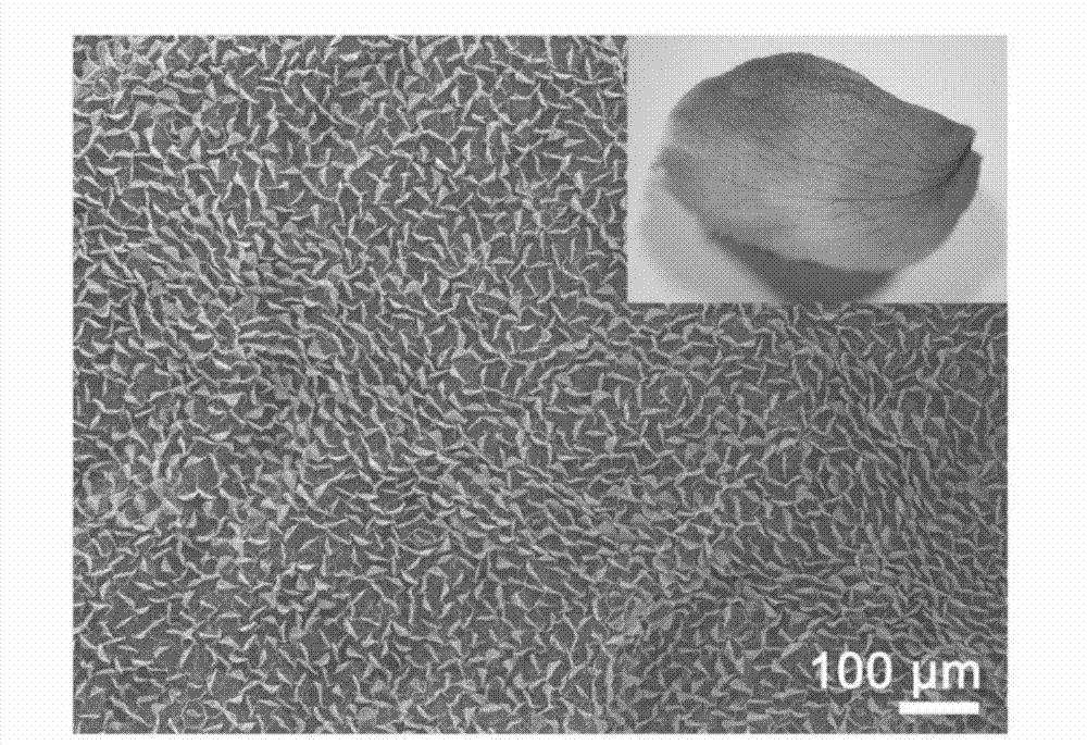
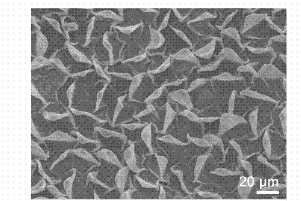
[0072] Select white rose petals and dry them as a whole or dry them naturally. The individual petals of dry white roses appear yellowish (the optical photo of which is shown in figure 1 shown in the middle inset), and its scanning electron microscope (SEM) shows that the surface of the petals is composed of some microsheet-like protrusions similar to flat shells, such as figure 1 As shown, the microsheet-like structure is uniformly distributed in a wide range. figure 2 For SEM with a slightly larger multiple, it can be seen that the spacing between the microstructures is 20-40 microns. When in contact with water droplets, it is easy to fo...

Embodiment 2

[0088] An argon ion laser was used as the excitation light source, the Raman spectrometer model used was JOBIN YVONT64000, the incident wavelength was 514 nm, and the laser power was 10 mW. The Raman scattering spectra of rhodamine 6G (R6G) molecular aqueous solutions with different concentrations were tested under a focusing microscope system. When the molecular concentration reaches 10 -9 When moles per liter, the test signal is still stable, that is, the detection limit concentration can reach 10-9 moles per liter. Compared with solid samples, the signal enhancement reaches 10 8 times. like Figure 13 shown. First put 10 -9 Mole per liter of liquid drops on the surface of 1 square centimeter dry rose petals steamed with silver, and as the water solvent volatilizes, R6G molecules are concentrated in a small area. 15 different test positions were selected in this local area, including 8 points on the top of the microstructure and 7 points on the side grating. Raman sig...


PUM

PropertyMeasurementUnit
thicknessaaaaaaaaaa
contact angleaaaaaaaaaa
angleaaaaaaaaaa

Abstract

The invention belongs to the technical field of ultra-sensitive test analysis, and particularly relates to an enhanced Raman detection substrate which can be in mass production and is wide in application, efficient, environment-friendly and based on a natural biology super-hydrophobic structure surface and a preparation method thereof. The PVD (physical vapor deposition) and a self-assembly or chemical plating and coating film technology are utilized to cover a precious metal layer on the natural biology super-hydrophobic surface to form rough and hydrophobic surface enhanced Roman detection hot spot area so that the enhanced Raman detection substrate is formed. The natural super-hydrophobic structure surface is plant pedals, plant leaves or inset wings. The precious metal is gold or silver and the thickness of the precious metal layer ranges from 5 nm to 50 nm. The hydrophobic SERS (surface enhanced Raman scattering) detection substrate has high sensitivity, and the detection limit can reach 10-9. The enhanced Raman detection substrate draws materials from the natural hydrophobic biology surface, so that the sensitivity is high, the performance is stable, the cost is low, and the pollution to the environment is prevented.

Description

technical field [0001] The invention belongs to the technical field of ultra-sensitive testing and analysis, and in particular relates to an enhanced Raman detection substrate based on a natural biological super-hydrophobic structure surface capable of large-scale production, wide application, high efficiency, and environmental protection and a preparation method thereof. Background technique [0002] Surface-enhanced Raman scattering (SERS) is an important technique in Raman spectroscopy. As an important means of trace analysis and detection, surface-enhanced Raman scattering (SERS) spectroscopy can provide direct fingerprint information of molecular vibration spectra, and has been widely studied and applied since it came out in 1974. SERS detection limit can reach femtograph (10 -15 ) level, making single-molecule detection possible and SERS detection technology can only enhance target molecules or groups in an extremely complex system, avoiding the interference of impuri...

Claims


Application Information

Patent Timeline
Patent Type & Authority Applications(China)
IPC IPC(8): G01N21/65C23C14/14C23C16/06
Inventor 徐彬彬孙洪波陈岐岱张然夏虹刘学青王磊
Owner JILIN UNIV
Why Eureka
Try Eureka