Method for preparing animal derived implantable medical biomaterial

一种动物源性、生物材料的技术,应用在医用生物材料领域,能够解决破坏三维结构、影响材料孔隙率、难以制备厚度和适应不同力学强度要求组织修复产品等问题,达到简化工艺流程、降低工艺耗时的效果

Active Publication Date: 2013-09-04
BEIJING BIOSIS HEALING BIOLOGICAL TECH
View PDF5 Cites 31 Cited by
  • Summary
  • Abstract
  • Description
  • Claims
  • Application Information

AI Technical Summary

Problems solved by technology

There are a variety of decellularization and virus inactivation methods reported, but most of them cannot completely remove all animal-derived DNA components, and most of them are time-consuming and require the use of various organic and high-strength acid-base solvents, resulting in decellularized SIS The destruction of the active extracellular matrix components in the matrix material, and the residue of harmful solvents will lead to cytotoxicity, thereby affecting the effect of tissue repair
In addition, most decellularization processes do not have effective measures to control endotoxin residues
The molding process is another technical difficulty in the preparation of acellular SIS matrix materials. The circumference of the small intestine is only 6-8cm, and the thickness of the submucosa of the small intestine is less than 0.1mm. It is difficult to prepare tissue repair products with different sizes, thicknesses and different mechanical strength requirements.
Cook Biotech Incorporated adopts the vacuum pressing method, which will compress the spatial structure of the decellularized SIS matrix material, destroy the natural three-dimensional structure of ECM, and affect the porosity of the material

Method used


Image

  • Method for preparing animal derived implantable medical biomaterial
  • Method for preparing animal derived implantable medical biomaterial
  • Method for preparing animal derived implantable medical biomaterial

Examples


Embodiment 1

[0031] Example 1: Preparation of decellularized porcine small intestinal submucosa matrix material

[0032] 1. Pretreatment separation and initial washing of animal tissue materials

[0033] The small intestine tissue of freshly slaughtered pigs was taken and cleaned, the submucosa of the small intestine was separated, and rinsed three times with water for injection.

[0034] 2. Virus inactivation

[0035] Use low-concentration peracetic acid-ethanol solution method to inactivate the virus. This step is carried out in a constant temperature ultrasonic cleaner that can shake the cleaning tank, wherein the volume percentage of peracetic acid is 0.05~0.2% (preferably 0.1%), The inactivation time is 1~2h (preferably 1h), the cleaning tank oscillation frequency is 30~600rpm (preferably 100~300rpm, more preferably 200rpm), the ultrasonic frequency is 20~80KHZ (preferably 20~50KHZ, more preferably 35~50KHZ, the most is 45KHZ), the temperature range is 4~40℃, and then wash 2~5 times...

Embodiment 2

[0044] Example 2: Physical and chemical properties, histology, growth factors and biological performance detection of the decellularized small intestine submucosa matrix material prepared in Example 1

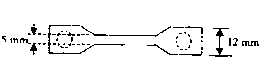
[0045] 1. The physical properties of the prepared 8-layer material were tested, and the test items included suture retention, tensile strength, burst strength and porosity.

[0046] 1) Suture retention test: Method: Use 2-0 surgical sutures or stainless steel wires of the same diameter to suture 2 mm inside the edge of one end of the 8-layer material, and fix the suture or stainless steel wire with the other end of the 8-layer material. On the tension meter, stretch at a speed of 20mm / min until the suture point is torn, and record the tension when the suture point is torn. Three batches of samples were tested according to the above method. Results: The suture tensile strength was greater than or equal to 5±0.5N.

[0047] 2) Tensile strength test method: method: use a tensile ...

Embodiment 3

[0065] Example 3: Preparation of decellularized porcine dermal matrix material

[0066] The dermal tissue of a freshly slaughtered pig is taken as a raw material, and the preparation method is the same as in Example 1.


PUM


Abstract

The invention provides a method for preparing an animal derived implantable medical biomaterial. The method comprises the following steps of: pretreatment, separation and primary cleaning of an animal tissue material; virus inactivation; decellularization; sodium chloride treatment; formation; packing and sterilization. According to the animal derived decellularized extracellular cell matrix (ECM) material prepared by using the method, animal derived cell components and DNA components are completely removed, meanwhile, the components and the three-dimensional structure of the natural ECM are completely kept, active growth factors for promoting tissue regeneration can be induced, residues of endotoxin, organic solvent and toxic solvent are avoided, and products with different sizes, thicknesses and mechanical strengths can be formed according to different indications.

Description

technical field [0001] The invention relates to the technical field of medical biomaterials, in particular to a method for preparing animal-derived implantable medical biomaterials. Background technique [0002] Defects and partial or total loss of functions of certain tissues or organs of the body caused by various diseases and traumas are one of the main hazards faced by human health. Research and development of ideal materials for tissue repair has always been an important topic in the fields of medicine, bioscience, and material science. At present, the medical biomaterials widely used clinically for tissue repair are mainly non-absorbable artificial materials, including polymer materials (such as polypropylene, polytetrafluoroethylene, polylactic acid, polyglycolic acid, silica gel, etc.), metal materials ( Such as stainless steel, titanium metal and its alloys, etc.), inorganic materials (such as bioactive ceramics, hydroxyapatite, etc.), composite materials (carbon ...

Claims


Application Information

Patent Timeline
Patent Type & Authority Applications(China)
IPC IPC(8): A61L27/36
CPCA61L27/3604A61L27/3629A61L27/3633A61L27/3687A61L27/3691
Inventor 赵博王振军
Owner BEIJING BIOSIS HEALING BIOLOGICAL TECH
Why Eureka
Try Eureka