Medical biological tissue structure and preparation method and special device of medical biological tissue structure

A technology of biological tissue and special equipment, which is applied in the field of tissue and organ manufacturing, can solve the problems of inability to meet the requirements of complex tissue and organ precursor manufacturing, single cells cannot be accurately positioned, coded, and the randomness of cell concentration is large, and it can achieve quantitative spraying technology Maximization and accuracy, elimination of mutual interference, and convenient manufacturing effects

Inactive Publication Date: 2014-07-02
TSINGHUA UNIV
View PDF14 Cites 12 Cited by
  • Summary
  • Abstract
  • Description
  • Claims
  • Application Information

AI Technical Summary

Problems solved by technology

[0012] This kind of design is simple and reliable, but the fixed multi-nozzle forming system has the following disadvantages: 1) low-temperature deposition manufacturing requires a low-temperature environment of minus 40 degrees, which will adversely affect the survival rate of cells, and cryopreservation agents need to be added , the process is more complex
2) The original system only has two nozzles, which can use two kinds of materials, but complex tissues and organs contain a variety of materials and cells, and the existing equipment cannot meet the requirements for the manufacture of complex tissue and organ precursors
3) The distribution concentration of cells inside the forming body is not uniform, the level of cell concentration is random, and a single cell cannot be accurately positioned and coded
4) It is impossible to spray the side surface of the forming body. The motor-assisted rapid prototyping nozzle needs to be installed vertically. If it is installed horizontally, the slurry will drip under its own gravity after being extruded from the nozzle, and cannot be sprayed on the forming body. side surface attachment

Method used


Image

  • Medical biological tissue structure and preparation method and special device of medical biological tissue structure
  • Medical biological tissue structure and preparation method and special device of medical biological tissue structure
  • Medical biological tissue structure and preparation method and special device of medical biological tissue structure

Examples


Embodiment 2

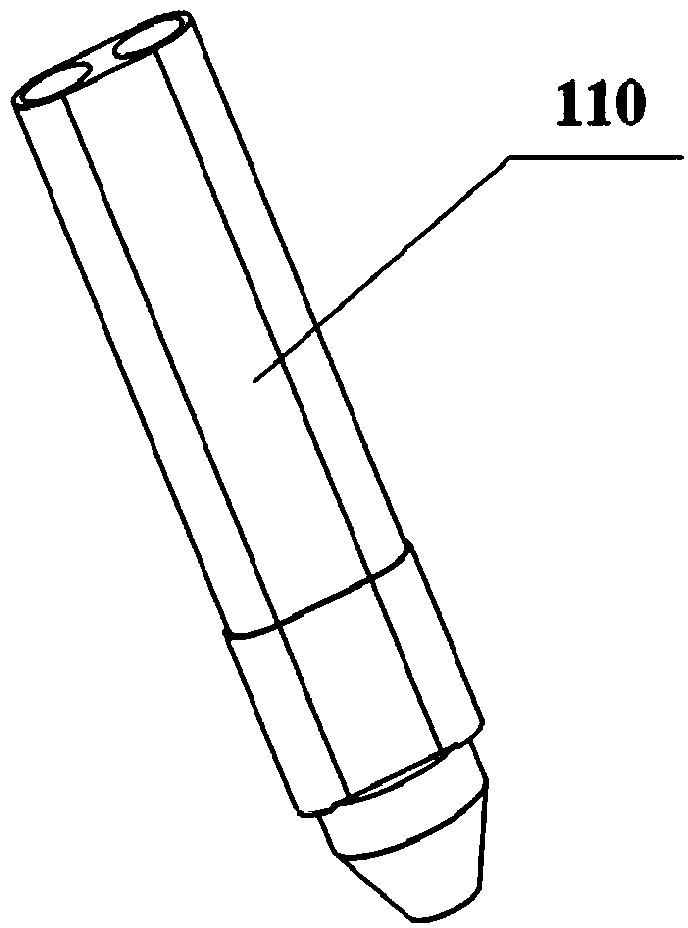
[0074] Embodiment 2: The natural polymer solution and the cross-linking agent are sodium alginate solution and calcium chloride solution. The concentration of sodium alginate solution is 5% (w / v), and the concentration of calcium chloride solution is 1% (w / v). Mix the endothelial cells into the sodium alginate solution at a density of 1 x 10 5 individual / mL. The sodium alginate solution and the calcium chloride solution containing endothelial cells are respectively loaded into two different component syringes of the hollow tube forming nozzle 110 for standby.

[0075] Cardiomyocytes and Schwann cells were extracted from patients. The above cells and sodium alginate solution were mixed. The cardiomyocyte density was 1 x 10 6 cells / mL, the Schwann cell density was 1×10 4 individual / mL. Fill the cell-containing polymer solutions into corresponding syringes for later use.

[0076] PLGA is dissolved in tetraethylene glycol solution to make a solution with a concentration of ...

Embodiment 3

[0085] Example 3: The natural polymer solution and the cross-linking agent are calcium hydrogen phosphate dihydrate (DCPD) and calcium hydroxide (1M disodium hydrogen phosphate) solution. The weight percentages of DCPD and calcium hydroxide in 1M disodium hydrogen phosphate solution are 20% and 10% (w / v) respectively. Put DCPD and calcium hydroxide solution into two different component syringes of the hollow tube forming nozzle 110 for standby.

[0086] Osteoblasts and adipose stem cells are extracted from patients. Mix the above cells with 5% (w / v) gelatin (PBS) solution. The density of osteoblasts was 1×10 5 cells / mL, the density of adipose-derived stem cells was 1×10 2 individual / mL. Add endothelial cell growth factor (10ng / mL) to fill the cell-containing polymer solutions into corresponding syringes for later use.

[0087] Dissolving PU in tetraethylene glycol solution to make a solution with a concentration of 10% (w / v), put it in the spray solution syringe of protec...

Embodiment 4

[0095] Example 4: The natural polymer solution and the cross-linking agent are polylactic-glycolic acid (PLGA) solution and water, respectively. Wherein the weight percent of PLGA in 1.4 dioxane is 20% (w / v). The PLGA solution and water are respectively loaded into two different component syringes of the hollow tube forming spray head 110 for standby.

[0096] Prepare 5% (w / v) gelatin (PBS) and 1% (w / v) fibrinogen solution and mix them, add 0.01% (w / v) heparin and osteoblast growth factor respectively, and put them into corresponding syringes for later use.

[0097] Dissolving PU in tetraethylene glycol solution to make a solution with a concentration of 20% (w / v), put it in the spray solution syringe of protective film nozzle 101 for subsequent use.

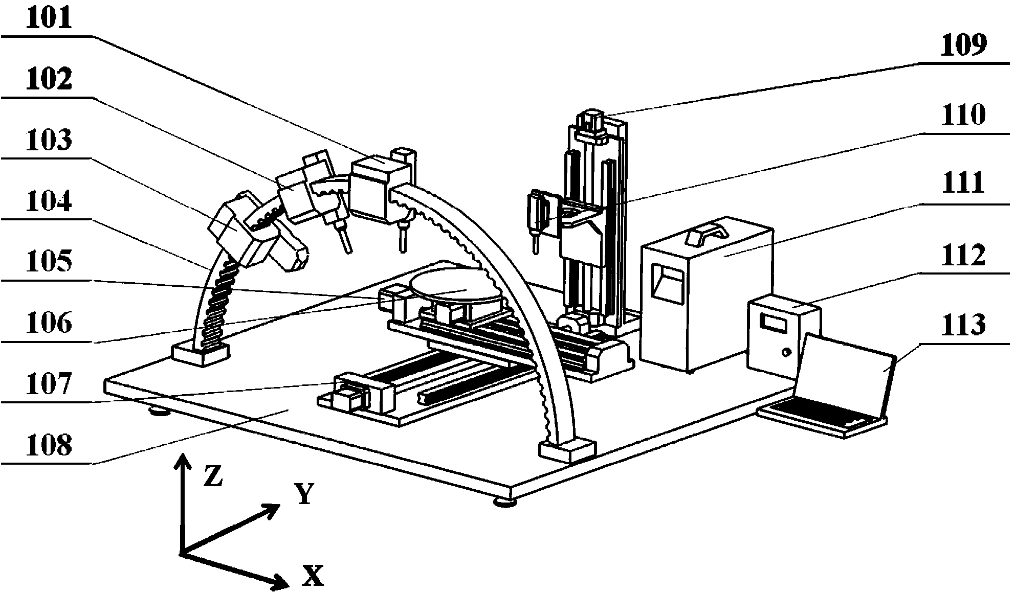
[0098] Driven by the X-direction motion device 105 and the Y-direction motion device 107, the rotary forming table 106 moves to the set position below the hollow tube forming nozzle, and the hollow tube forming nozzle 110 extru...


PUM


Abstract

The invention relates to a medical biological tissue structure and a preparation method and a special device of the medical biological tissue structure, and belongs to the technical field of biology, medical treatment and medical instruments. The medical biological tissue structure comprises a hollow pipe, a functional layer and a synthetic macromolecule protective film, wherein the hollow pipe is made of biological materials containing cells or being free of the cells, and the functional layer is attached to the hollow pipe and contains or does not contain the cells. The medical biological tissue structure, the preparation method and the special device are based on the attachment cross-linking and solidity principle, under control of a computer, the hollow pipe is firstly extruded out through a special spraying nozzle, then the biological materials are sprayed and attached to the hollow pipe to form the functional layer and accumulated layer by layer to form a composite molding body, and finally a synthetic macromolecule solution is sprayed to the outer surface of the composite molding body to form the protective film. According to the set molding steps, a three-dimensional structure body which contains synthetic macromolecule materials, the cells and the natural biological materials is finally manufactured, has the space complex shape and is of the gap structure is finally manufactured. The medical biological tissue structure can be molded at the normal temperature, the process is simple, the cell survival rate is high, distribution is even and controllable, and the good mechanical property and the good biological property are achieved.

Description

technical field [0001] The invention belongs to the technical field of tissue and organ manufacture, and in particular relates to a medical biological tissue structure, its preparation method and special equipment. Background technique [0002] Large tissues (such as bone, cartilage, breast, muscle, skin) and internal organs (such as heart, liver, spleen, lungs, kidneys, etc.), which are important components of the human body, are damaged and Dysfunction seriously endangers human health and quality of life. Traditional clinical methods still have many limitations, especially the inability to reconstruct the structure of controlled distribution of cells in vitro. The cross fusion of manufacturing science and life science has given birth to the controlled assembly technology of cells, which has opened up a new way for the direct construction of organ substitutes with new functions in vitro. [0003] Low-temperature deposition manufacturing process (LDM) is a new process deve...

Claims


Application Information

Patent Timeline
Patent Type & Authority Applications(China)
IPC IPC(8): A61F2/02
CPCA61F2/02
Inventor 王小红刘利彪
Owner TSINGHUA UNIV
Why Eureka
Try Eureka