
Cell tissue resonance Raman spectroscopy scanning imaging method

A technology of cell tissue and Raman spectroscopy, applied in the field of imaging analysis, can solve the problem of limited number of Raman spectra, achieve the effects of shortening detection time, simple operation, and enhancing Raman signal intensity

Inactive Publication Date: 2014-10-22
TECHNICAL INST OF PHYSICS & CHEMISTRY - CHINESE ACAD OF SCI
View PDF3 Cites 9 Cited by
  • Summary
  • Abstract
  • Description
  • Claims
  • Application Information

AI Technical Summary

Problems solved by technology

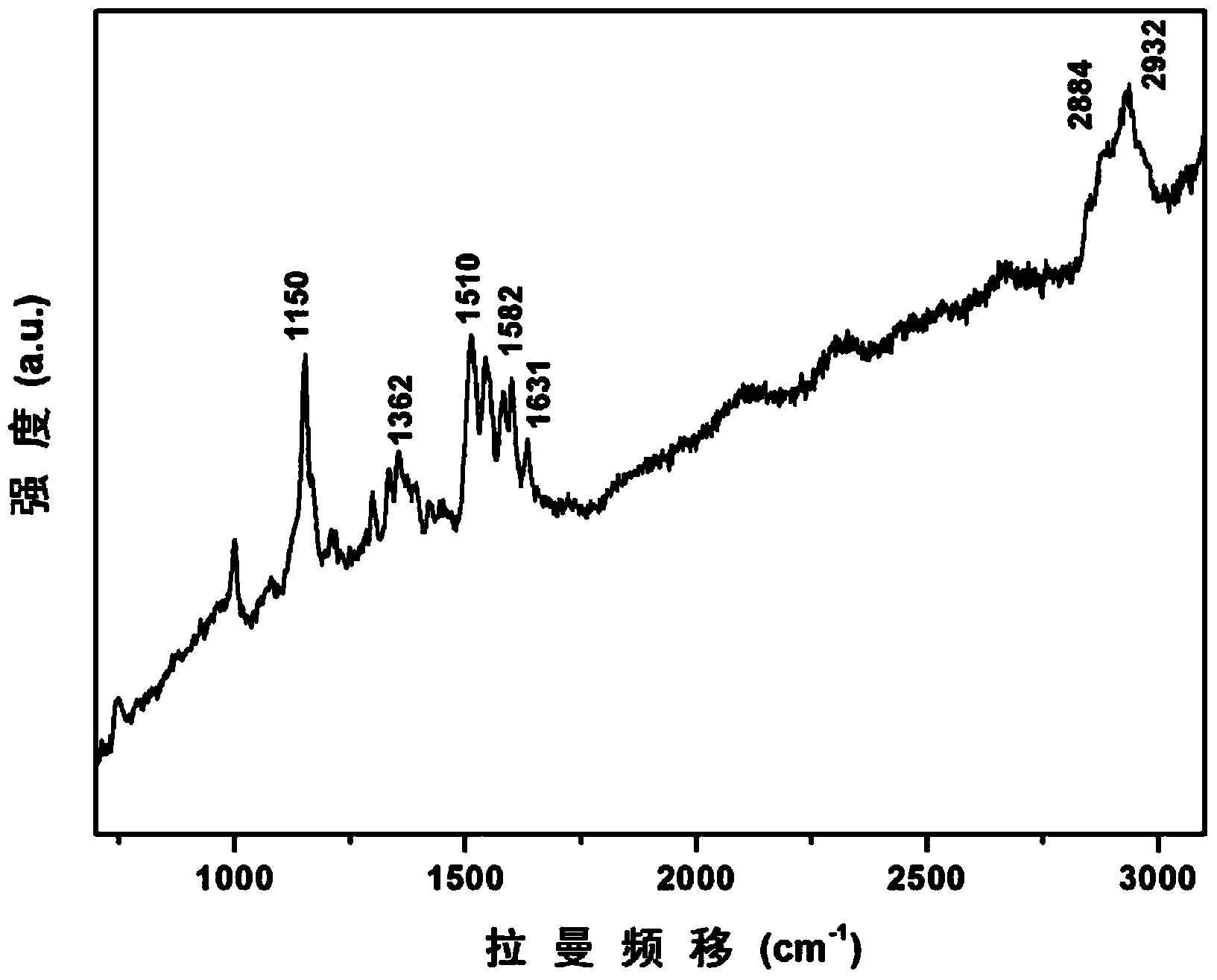
Although the biological macromolecules that make up the cell tissue have a resonance response in the blue-green light band, the Raman signal of the sample can be strengthened through the resonance effect by selecting a laser with a suitable wavelength as the excitation light source, and the measurement time can be shortened without destroying the activity of the sample. time, but the scanning time of the whole area is still more than 1 hour, and the number of Raman spectra provided per unit time is limited

Method used


Image

  • Cell tissue resonance Raman spectroscopy scanning imaging method
  • Cell tissue resonance Raman spectroscopy scanning imaging method
  • Cell tissue resonance Raman spectroscopy scanning imaging method

Examples


Embodiment 1

[0040] A resonance Raman spectroscopy rapid scanning imaging method for identifying cell tissues, the specific steps of which are as follows:

[0041] (1), the brain cell tissue samples were frozen;

[0042] Will (5x5x2mm 3 ) The brain tissue block was placed flat in a small stainless steel metal box (box size: 20mmx20mmx5mm), and the box was slowly and flatly placed in a small cup filled with liquid nitrogen. When the bottom of the box touched liquid nitrogen, it began to vaporize and boil. At this time, keep the small box in place and do not immerse it in liquid nitrogen, and the tissue will quickly freeze into blocks in about 10-20 seconds. After the frozen block is made, quickly wrap it with plastic film, and immediately put it in the refrigerator for storage. The temperature of the refrigerator is set at -70°C.

[0043] (2) Use a cryostat to slice the aforementioned tissue frozen block. Keep the fat-containing portion of the tissue perpendicular to the incision. Slici...

Embodiment 2

[0049] A resonance Raman spectroscopy rapid scanning imaging method for identifying cell tissues, the specific steps of which are as follows:

[0050] (1), the breast tissue sample is frozen;

[0051] Will (7x7x4mm 3 ) mammary gland tissue blocks were placed flat in a small copper box (box size: 20mmx20mmx5mm), and the box was slowly and flatly placed in a small cup filled with liquid nitrogen. When the bottom of the box touched liquid nitrogen, it began to vaporize and boil. At this time, keep the small box in place and do not immerse it in liquid nitrogen, and the tissue will quickly freeze into blocks in about 10-20 seconds. After the frozen block is made, quickly wrap it with a plastic film, and immediately put it in the refrigerator for storage. The temperature of the refrigerator is set at -120°C.

[0052] (2) Use a cryostat to slice the aforementioned tissue frozen block. Keep the fat-containing portion of the tissue perpendicular to the incision. Slicing is carried...

Embodiment 3-8

[0056] Same as Example 1, the difference is that the brain tissue samples in Example 1 are replaced by glioma tissue, breast tissue, gastric mucosal tissue, rectal tissue, ovarian tissue, and uterine tissue, and the obtained results are the same as those in Example 1. approximate.


PUM

PropertyMeasurementUnit
thicknessaaaaaaaaaa

Abstract

The invention discloses a cell tissue resonance Raman spectroscopy scanning imaging method which comprises the following step of: carrying out (1) freezing treatment and (2) sample preparation and scanning imaging on a cell tissue. The cell tissue resonance Raman spectroscopy scanning imaging method is easy and fast to operate, can fast carry out qualitative detection on a tissue sample, effectively enhances the Raman detection sensitivity, inhibits the fluorescence disturbance, enhances the Raman signal intensity, ensures the spectroscopy imaging quality, shortens the detection time and visually shows the distribution of characteristic components of the tissue sample in a clear image mode.

Description

technical field [0001] The invention relates to the technical field of imaging analysis. More specifically, it relates to a rapid scanning imaging method of cell tissue resonance Raman spectroscopy. Background technique [0002] At present, the imaging methods of tissues and cells in medicine mainly use means such as magnetic resonance imaging (MRI), X-ray scanning, computed tomography (CT) or positron emission tomography (PET). [0003] Among the above detection techniques, X-ray scanning and computed tomography (CT) images are scanned by X-rays, so they have certain radioactivity; magnetic resonance imaging (MRI) takes a long time, and the completion time varies depending on the scanning site. The change usually takes more than 1 hour, while the scan time of positron emission tomography (PET) is shorter, about 20-30 minutes, but the preparation time before scanning is longer, and it takes about 1 hour. These testing devices are mostly deployed in hospitals for clinical t...

Claims


Application Information

Patent Timeline
Patent Type & Authority Applications(China)
IPC IPC(8): G01N21/65G01N1/28
CPCG01N21/65
Inventor 段宣明刘洁郑美玲
Owner TECHNICAL INST OF PHYSICS & CHEMISTRY - CHINESE ACAD OF SCI
Learn More