A rapid and sensitive method for the detection of small RNAs

A sensitive detection, small molecule technology, applied in biochemical equipment and methods, microbiological determination/inspection, etc., can solve the problems of not showing bands, background interference, long time, etc., to shorten the detection time and improve the detection sensitivity , The effect of shortening the detection time

Inactive Publication Date: 2017-03-29
SICHUAN UNIV
View PDF0 Cites 0 Cited by
  • Summary
  • Abstract
  • Description
  • Claims
  • Application Information

AI Technical Summary

Problems solved by technology

However, this method cannot display bands, so it cannot distinguish small RNAs with similar sequences, and it is difficult to detect multiple small RNAs
Guo Yaohui et al. established a biotin-labeled-labeled method in "Biotin-labeled probe-liquid phase hybridization-non-denaturing PAGE method for detecting non-coding small RNA" (International Journal of Laboratory Medicine, Volume 33, Issue 6, March 2012). Liquid-phase hybridization-non-denaturing polyacrylamide gel electrophoresis is a method for detecting non-coding small RNAs. However, this method has a large background interference problem because it cannot remove redundant probes, and it is easy to produce false positive results that affect judgment, and it is difficult to achieve sensitivity. , accurate detection
In terms of actual detection effect, in order to obtain a better detection signal, the probe concentration required by this method is as high as 10 umol / L; and it takes a long time, from hybridization to detection of signal, excluding the time required for electrophoretic separation and membrane transfer. It takes 5 hours

Method used


Image

  • A rapid and sensitive method for the detection of small RNAs
  • A rapid and sensitive method for the detection of small RNAs
  • A rapid and sensitive method for the detection of small RNAs

Examples


Embodiment 1

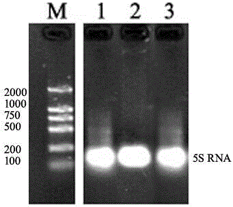
[0037] In this example, the detection of Osa-miR156 in rice leaves, let-7-a in mouse hepatocytes, and chicken blood was taken as an example.

[0038] The flow chart of the whole detection method is as follows: figure 1 shown.

[0039] In the process of realizing the object of the present invention, the following steps are usually included:

[0040] 1. Material selection: fresh animal and plant tissues, blood, or fresh -70°C frozen samples.

[0041] 2. Reagent configuration:

[0042] 1) DEPC treated water RNase free water: deionized water is treated by Millpore water processor, add 1 / 1000 DEPC water after treatment, sterilize under high temperature and high pressure for 30 minutes, and cool to room temperature;

[0043] 2) TBE: Tris: 108g; Na 2 EDTA·2H 2 O: 7.44g; boric acid: 55g, add 800ml of deionized water, stir to dissolve, add deionized water to 1L, store at room temperature;

[0044] 3) 10% APS: 1g ammonium persulfate, dissolved in 10ml double distilled water;

[...

Embodiment 2

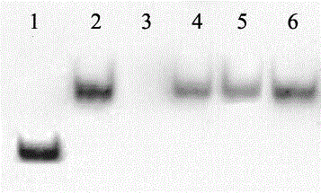
[0083] Detection sensitivity test

[0084] This embodiment is based on the method of Example 1. During liquid phase hybridization, Osa-miR156 in rice leaves with different concentrations was used for liquid phase hybridization and solid phase detection, and the detection sensitivity test was carried out. The results are as follows: Figure 4 as shown, Figure 4The concentrations of Osa-miR156 in bands 1-15 are 1. 10 fmol; 2. 5 fmol; 3. 2.5 fmol; 4. 1 fmol; 5. 0.5 fmol; 6. 0.25 fmol; 7. 0.1 fmol; 8. 0.05 fmol; 9. 0.025 fmol; 10. 0.01 fmol; 11. 0.005 fmol; 12. 0.0025 fmol; 13. 1 amol;

[0085] From Figure 4 It can be seen that this method can detect at least 0.005 fmol of miRNA (5aM=35fg=0.035pg), that is, band 11 can be clearly identified, and the sensitivity of this method is 10 times higher than that of the previous small RNA detection method.

Embodiment 3

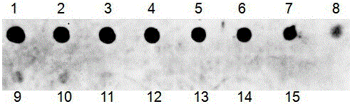
[0087] Small molecule RNA quantitative detection test

[0088] In this example, the quantification of miR156 in rice was taken as an example; the DNA form miD156 of miR156 with a series of concentration gradients (0.8-31.2 fmol) was hybridized with 0.1 pmol of biotin-labeled probe miD156*, and ChemiDoc XRS was used after color development The detection system carried out relative quantitative analysis. The concentration of miD156 used for hybridization was used as the X axis, and the chromogenic concentration was used as the Y axis to establish a coordinate curve. The results showed a good linear relationship between the two, y = 34.671x + 0.231, and R = 0.9902 in the linear regression equation. Band 7 is the hybridization of 1ug rice miRNA with 0.1pmol miD156* probe, the result is in the regression equation,

[0089] The result is as Figure 5 as shown, Figure 5 In , the miR156 concentrations of bands 1-7 were 1: 0.8fmol; 2:1.9fmol; 3: 3.9fmol; 4: 7.8fmol; 5: 15.6fmol; 6: ...


PUM


Abstract

The invention discloses a method for quickly and sensitively detecting small RNA, belonging to the field of molecular biology, comprising the following steps: (1) extracting and purifying the small RNA to be detected; (2) probe design: the probe uses biotin or ground (3) liquid-phase hybridization: hybridize the small molecule RNA to be detected with the probe in the buffer, and use exonuclease to excise the unhybridized excess single strand and then perform gel electrophoresis; (4) Membrane transfer: transfer the hybridization product on the gel after electrophoresis to a solid support (such as nylon membrane, cellulose membrane); (5) Antibody incubation: transfer the hybridization product obtained in step (4) The solid phase support and the specific antibody are incubated for hybridization; (6) signal detection. The method can detect at least 0.005 fmol of miRNA, and the sensitivity is at least 10 times higher than that of the prior art. The method has a fast detection speed and can realize high-throughput detection and quantitative detection.

Description

technical field [0001] The invention relates to the field of molecular biology, in particular to a method for rapidly and sensitively detecting small molecule RNA. Background technique [0002] MicroRNAs (miRNAs) are a class of non-coding endogenous RNAs that have been found in multicellular species and even unicellular species. MicroRNAs can reflect gene expression from transcription to translation levels. Therefore, in different tissues and There are great differences in the expression levels of different developmental stages, and there are also great differences among different small RNAs. A series of research data have shown that small RNAs (miRNAs) can regulate gene expression at the post-transcriptional level through complementary base repair mode, and can also regulate gene expression at the DNA level by promoting DNA methylation. Therefore, small RNAs play important roles in almost all biological aspects including physiological, biochemical, developmental and pathol...

Claims


Application Information

Patent Timeline
Patent Type & Authority Patents(China)
IPC IPC(8): C12Q1/68
CPCC12Q1/6804C12Q2525/207C12Q2563/131
Inventor 陈放李佛生毛强詹诚唐琳
Owner SICHUAN UNIV
Why Eureka
Try Eureka