


Quick Research
Generate reliable direction feasibility study reports for your R&D in just a few steps.

Technical Q&A
Discover and master advanced knowledge NOW. Basics, ideas, possibilities, all at once.

Find Solutions
As an expert in R&D theories, this can generate solutions to your technical problems instantly.

Evaluate Feasibility
Analyze your overall solution with one click, know your potential R&D risks in advance.

Monitor Landscape
Get weekly tech updates, stay abreast of the latest tech innovations and key insights.
Method for remotely delivering recombinant amniotic culture epithelial membrane
A membrane and epithelial technology, applied in the field of cell culture, can solve problems such as poor mastery, lack of medium, cumbersome process, etc., and achieve the effect of convenient remote delivery, maintaining cell viability, and good cell viability
- Summary
- Abstract
- Description
- Claims
- Application Information
AI Technical Summary
Problems solved by technology
Method used
Image

Examples
preparation example Construction
[0013] 1. Preparation of three-dimensional gelatin scaffolds:
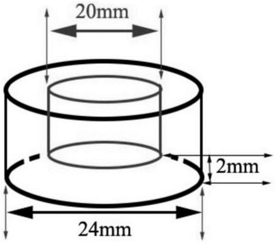
[0014] Prepare the gelatin sponge bottom pad: use a compass to draw a circle with a diameter of 32mm on the gelatin sponge, measure the height to 6mm, and cut the round pad to make the bottom pad;
[0015] Preparation of gelatin cylinder ring: use a compass to draw a circle with a diameter of 25mm on the gelatin sponge, measure the height to 10mm, cut off the cylinder, keep the wall thickness of the cylinder at 3mm, and cut off the rest with a circular stainless steel knife. Sterilize the prepared base pad and cylindrical ring with ethylene oxide for later use ( figure 1 with figure 2 ).
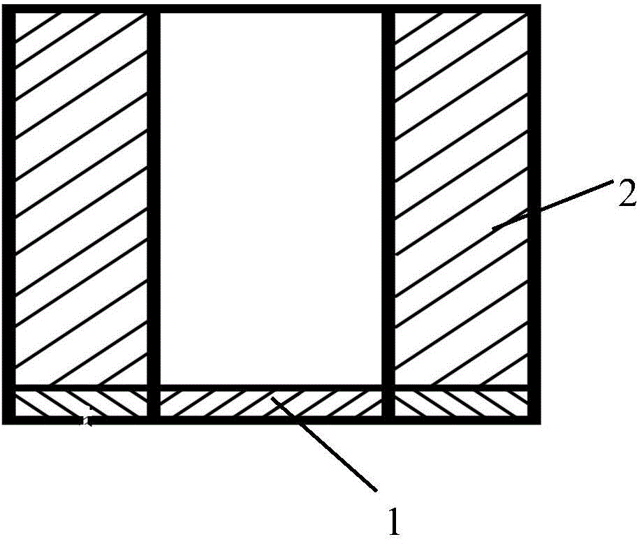
[0016] 2. Three-dimensional three-dimensional gelatin scaffold packaging cell membrane:
[0017] Put the sterilized gelatin sponge pad into the bottom of the 6-well culture plate, place the gelatin sponge ring on the edge of the culture well, take out the cultured cell membrane sleeve from the transwell, insert the epithelia...
Embodiment 1
[0022] After transporting for 48 hours, take out the cell membrane, add 0.25% trypsin-0.02% EDTA to half of the membrane, digest at 37°C for 10 minutes, centrifuge at 1500rpm / min for 5 minutes, add cell culture medium and centrifuge to resuspend to make a single cell suspension, take 10μl of cells Suspension, add an equal volume of 0.4% trypan blue solution, pipette and mix well, use a cell counter to count the proportion of living cells and the total cell amount, the total amount of cells is 6.26×10 5 , the proportion of living cells was 56%; the other half of the cell membranes were fixed with paraformaldehyde, embedded in paraffin, made paraffin sections, and stained with HE to observe the cell morphology and membrane structure. Attached to the homogeneous amniotic membrane surface, about 3-5 layers, the cells are closely arranged, the bottom layer cells are nearly cuboid, while the upper layer cells are relatively flat.
Embodiment 2
[0024] Prepare the three-dimensional gelatin scaffold according to the conventional method, put the sterilized gelatin sponge pad into the bottom of the 6-well culture plate, put the gelatin sponge ring on it, take out the cultured cell membrane from the transwell, and insert the epithelial side up into the gelatin sponge ring Inside, add nutrient solution to the sponge until it is saturated, cover the culture plate, seal the culture plate with a sealing film, put it in an insulated box and send it by express delivery.
[0025] In the traditional transportation method, after the cultured cell membrane is taken out, it is directly placed in a 60mm culture dish, a small amount of medium is added to cover the membrane, and the sealing film is sealed and then packed in a box for express delivery. Since the cell membrane cannot be fixed, it is very easy to cause the cell membrane or cells to fall off due to position changes during transportation, and the culture medium in the cultur...
PUM
| Property | Measurement | Unit |
|---|---|---|
| Diameter | aaaaa | aaaaa |
| Height | aaaaa | aaaaa |
| The inside diameter of | aaaaa | aaaaa |
Abstract
Description
Claims
Application Information
 - R&D Engineer
- R&D Manager
- IP Professional
- Industry Leading Data Capabilities
- Powerful AI technology
- Patent DNA Extraction
Browse by: Latest US Patents, China's latest patents, Technical Efficacy Thesaurus, Application Domain, Technology Topic, Popular Technical Reports.
© 2024 PatSnap. All rights reserved.Legal|Privacy policy|Modern Slavery Act Transparency Statement|Sitemap|About US| Contact US: help@patsnap.com