Method for detecting embryonic chromosome abnormality by virtue of blastochyle free DNA

A chromosomal abnormality and blastocoel fluid technology, applied in biochemical equipment and methods, microbial determination/inspection, etc., to achieve the effects of low probability, simple operation, and convenient material acquisition.

Inactive Publication Date: 2015-03-25
SUZHOU BASECARE MEDICAL DEVICE CO LTD
View PDF4 Cites 18 Cited by
  • Summary
  • Abstract
  • Description
  • Claims
  • Application Information

AI Technical Summary

Problems solved by technology

This kind of preimplantation chromosome screening by taking single cells at the cleavage stage or blastocyst stage is widely used in clinical practice, but there is no large amount of clinical data to prove that single-cell samples at the cleavage stage or blastocyst stage have a significant effect on the later development of embryos. Whether there is an impact, so it is necessary to find a safer detection marker that has no impact on embryos in clinical practice

Method used


Image

  • Method for detecting embryonic chromosome abnormality by virtue of blastochyle free DNA
  • Method for detecting embryonic chromosome abnormality by virtue of blastochyle free DNA
  • Method for detecting embryonic chromosome abnormality by virtue of blastochyle free DNA

Examples


Embodiment 1

[0059] Example 1 Mouse blastocoel fluid free DNA detection

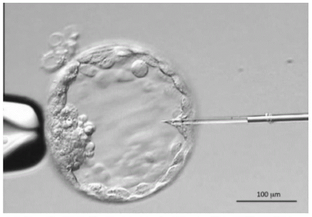
[0060] (1) Collection of blastocoel fluid

[0061] Acquisition and processing of mouse blastocoel fluid: the acquisition of mouse blastocoel fluid DNA and the acquisition of mouse single cells are the same mouse embryo, so as to facilitate the comparison of test results. In the micromanipulation laboratory, samples were taken by micropuncture technique at room temperature. In the process of aspirating blastocoel fluid DNA, avoid cytoplasm contamination and damage to trophoblast cells. The DNA sampling process of mouse blastocoel fluid is as follows: figure 1 shown.

[0062] (2) Target gene and primer design

[0063] Two genes, GAPH and TBCD, were used as detection genes, and primers were designed for quantitative analysis and detection. The sizes of the amplified fragments of GAPDH and TBCD were 100bp and 120bp.

[0064] Amplification primer sequences are as follows.

[0065] GAPDH:

[0066] P1 TGTCTCACC TTAT...

Embodiment 2

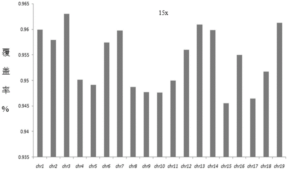
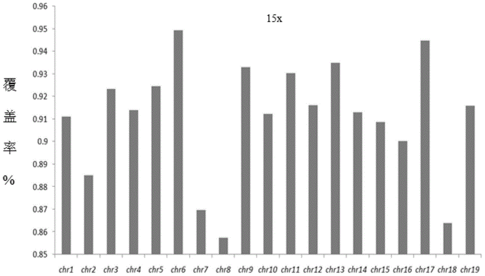
[0077] Comparison of mouse genome sequencing coverage (mouse embryo single cell and mouse blastocoel fluid DNA)

[0078] (1) Single cell isolation and blastocoel fluid DNA acquisition and processing:

[0079] Pick 10 mouse embryo single-cell samples and 10 mouse blastocoel fluid samples, and each mouse embryo single-cell sample and mouse blastocoel fluid sample correspond one-to-one, and come from the same mouse embryo.

[0080] a. Isolation and processing of mouse embryo single cells: under a microscope, single cells were isolated by laser drilling technology, and the isolated single cells were placed in a 200 μl PCR tube containing less than 2 μl of PBS, and water was added to a final volume of 9 μl. Add 1 μl of freshly prepared cell lysis buffer to the PCR tube and mix well. The resulting mixture was incubated: 50°C for 1h, 99°C for 4min, 15°C hold, cooled on ice, and centrifuged.

[0081] b. Acquisition and processing of mouse blastocoel fluid: the acquisition of mouse b...

Embodiment 3

[0098] Chromosomal Abnormality Analysis of Mouse Genome Sequencing (Mouse Embryo Single Cell and Mouse Blastocoel Fluid DNA)

[0099] (1) The purpose of this experiment is to analyze that blastocoel fluid can be used for chromosomal abnormality analysis instead of single-cell samples. Two gene knockout mice from our laboratory were selected, one of which was chromosomal deletion and the other was chromosomal duplication. Each mouse was subjected to blastocoel fluid acquisition and embryonic single cell isolation. After the genomic DNA was amplified and the amplified product was qualified, fragmentation processing and library construction were performed. The experimental procedure was the same as in Example 2.

[0100] (2) On-machine sequencing. The quality inspection of the samples constructed in the above experiments was carried out, and the quality inspection was qualified, and the Ion Proton system was used for whole genome sequencing.

[0101] (3) Biological information ...


PUM


Abstract

The invention relates to a method for detecting embryonic chromosome abnormality by virtue of blastochyle free DNA. The method comprises the following steps: acquiring blastochyle free DNA, detecting the blastochyle DNA, carrying out whole genome amplification of the free DNA, analyzing a product of the whole genome amplification, implementing fragmenting treatment on genome DNA, carrying out quantitative analysis and fragment size analysis on fragmented target DNA, constructing a library, sequencing by virtue of a computer and analyzing biological information. By virtue of high-throughput sequencing, the method disclosed by the invention can be used for overcoming shortcomings of a conventional DNA analysis method which is merely used for researching partial region of a single cell genome, and is capable of completely analyzing the genetic information of the single cell genome; the method is simple and convenient to operate, time-saving and efficient; meanwhile, by using the blastochyle free DNA as a detection sample, the method is convenient and safe to sample, so that the probability of later embryonic development abnormality is reduced and embryo is protected from being influenced in later development.

Description

【Technical field】 [0001] The invention relates to the field of molecular cell biology, in particular to a method for detecting embryo chromosomal abnormality by using free DNA of blastocoel fluid. 【Background technique】 [0002] Some specific biomarkers produced in organisms, such as proteins, peptides, lipids, RNA, DNA and their variants and modifications, can be applied to the diagnosis, prognosis and treatment of diseases. These biomarkers can exist in a variety of body fluids, including blood, plasma, serum, breast milk, ascites and urine, etc., and have been applied to cancer (prostate cancer, breast cancer, liver cancer, pancreatic cancer, ovarian cancer, esophageal cancer, Colon cancer, etc.), similar cancers, B-cell lymphoma, Parkinson's disease, Alzheimer's disease, sexual prion disease and other related diseases and genetic diseases. At present, it has been found that the peripheral blood of pregnant women contains fetal circulating cell-free DNA, and the fetal ce...

Claims


Application Information

Patent Timeline
Patent Type & Authority Applications(China)
IPC IPC(8): C12Q1/68
CPCC12Q1/6869
Inventor 梁波孔令印冒艳杨晴吴小娟
Owner SUZHOU BASECARE MEDICAL DEVICE CO LTD
Why Eureka
Try Eureka