
A two-dimensional light scattering static cytometer method and device

A light scattering and cytometer technology, applied in the fields of biotechnology, medical devices, environment and safety, and biomedicine. Sophisticated, broadly applicable effects

Active Publication Date: 2017-10-17
SHANDONG UNIV
View PDF7 Cites 0 Cited by
  • Summary
  • Abstract
  • Description
  • Claims
  • Application Information

AI Technical Summary

Problems solved by technology

[0006] (1) Due to the limitation of the diffraction limit, the highest resolution of the traditional optical microscope for cell and particle analysis is about 300nm. At this time, it has higher requirements for the numerical aperture of the objective lens. If you need to use 100 times the oil Immersion objective lens;
[0007] (2) Traditional cell analysis devices such as flow cytometers analyze cell size through forward scattered light information, but there are problems of low analysis accuracy and inability to clearly identify cell size;
[0008] (3) Since the excitation light path of the flow cytometer is fixed, it is generally perpendicular to the axis of the cell suspension, which requires that the cells must not deviate from the axis when flowing through the laser focus area, and must not aggregate into clusters, Block the pipeline, otherwise the beam cannot accurately irradiate the center of the cell, resulting in unstable signal and affecting the accuracy of the measurement results
This requires complex and expensive fluid control techniques, such as hydrodynamic focusing, which are not suitable for most laboratories;
[0009] (4) The use of nozzles with different apertures and changing the liquid flow velocity will affect the cell analysis and sorting effect, and a delay time is required from the parameter measurement to the pulse charging through logic selection, and the accurate determination of the delay time also affects the sorting quality. The key, which has caused the complexity of the operation and use of flow cytometer;
[0010] (5) The traditional scanning flow cytometer only measures the azimuth one-dimensional light scattering. Due to the arbitrary shape of the cells and the problem of the axial direction of the cells, a lot of information will be lost in the one-dimensional scattering spectrum obtained at a fixed angle;
[0011] (6) Flow cytometry detection requires fluorescent staining or labeling of the tested cells with special dyes, which will cause certain damage to the cells and affect cell viability;
[0012] (7) Electromagnetic field cell sorting of traditional flow cytometry will have a certain impact on living organisms
These measurement methods usually require a complex optical path system and have high requirements on the size and shape of the sample to be measured, which limits its application to a certain extent, especially in the measurement of the refractive index of cells and particles
[0014] In short, the existing cytometers have certain limitations, and there is an urgent need for a new type of two-dimensional light scattering static cytometer device and method of use.

Method used


Image

  • A two-dimensional light scattering static cytometer method and device
  • A two-dimensional light scattering static cytometer method and device
  • A two-dimensional light scattering static cytometer method and device

Examples


Embodiment 1

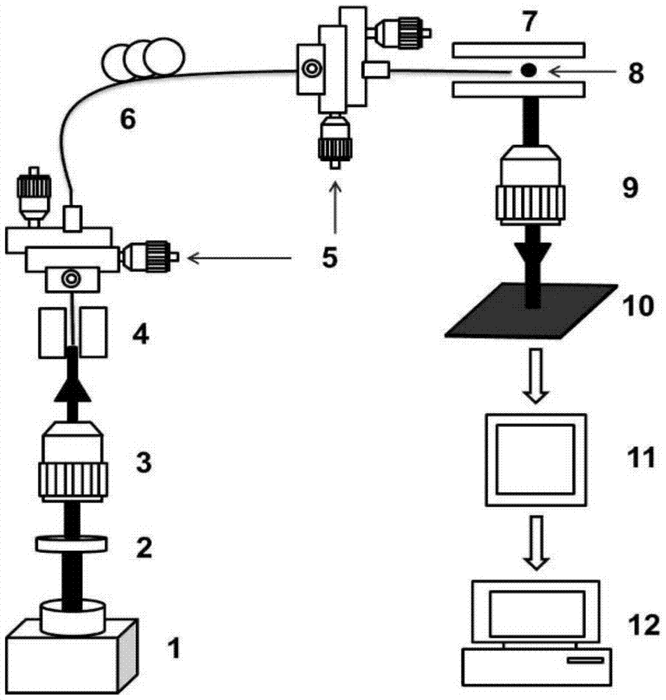
[0063] The structural principle of the device of the present invention is as figure 1 As shown, yeast cells were selected as the tested samples for nanoscale analysis of cell size.

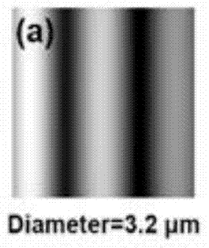
[0064] According to the Mie scattering theory, yeast cells are assumed to have spherical particles with different diameters, the refractive index of the cells is 1.42, the incident light wavelength is 532nm, and the refractive index of the surrounding medium and aqueous solution is 1.334. The nanometer resolution single cells and particles provided by the present invention The angle range of the scattering pattern detected in the analyzed two-dimensional light scattering static cytometer device is 79-101 degrees, so the theoretical simulation also selects the same angle range, and then simulates the two-dimensional light scattering of a large number of yeast cell models with known diameters Pattern 11, such as Figure 3(a)-Figure 3(j) shown.

[0065] Preparation of the single-cell thin layer of ...

Embodiment 2

[0070] The structural principle of the device of the present invention is as figure 1 As shown, microspheres with average diameters of 3.87 μm (standard error 0.25 μm) and 4.19 μm (standard error 0.27 μm) were selected as the tested samples for submicron level analysis of particle size.

[0071] The refractive index of theoretically simulated microspheres is 1.42, the incident light wavelength is 532um, the refractive index of the surrounding medium and aqueous solution is 1.334, and the angle range is 79-101 degrees. A large number of microsphere models with known diameters are simulated by Mie scattering theory. Dimensional light scattering pattern11.

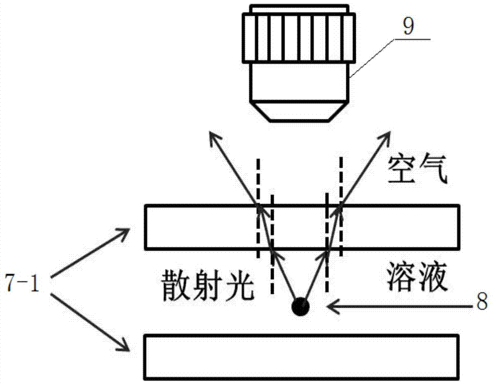
[0072] The measured microsphere solution is diluted to ensure that there is a small amount of free microspheres in the observation field of view of the ten times objective lens 9. Use a pipette to draw a small amount of microsphere solution each time, drop it on the center of slide glass 7-1 with a cover glass placed at each...

Embodiment 3

[0077] The structural principle of the device of the present invention is as figure 1 As shown, yeast cells with a refractive index of 1.42 (incident wavelength of 589 nm) and microspheres with a refractive index of 1.59 (incident wavelength of 589 nm) were selected as tested samples for refractive index analysis.

[0078] According to the Mie scattering theory, 45 two-dimensional light scattering patterns 11 of the yeast model with a refractive index of 1.42, an incident light wavelength of 532nm, a refractive index of the surrounding medium and aqueous solution and different diameters (diameters ranging from 2.8 μm to 7.2 μm) were obtained by simulation. μm, with a step size of 0.1 μm), scan the 45 scattering patterns horizontally to obtain the change curve of the grayscale image; perform FFT on the grayscale curve obtained by scanning, and a typical main peak will appear in the FFT curve, and make statistics on it Analysis, the linear equation of the yeast cell peak frequen...


PUM

PropertyMeasurementUnit
diameteraaaaaaaaaa
diameteraaaaaaaaaa
diameteraaaaaaaaaa

Abstract

The invention discloses a two-dimensional light scattering static cytometer method and device, including a laser light source, the laser light source is sequentially connected with a neutral density filter, an optical lens and an optical fiber coupling system, and the optical lens and the optical fiber coupling system adjust the laser , so that the optical fiber is used as a probe to excite the static cells and particles on the static cell and particle sample placement device, the static cell and particle sample placement device is connected with the two-dimensional light scattering pattern detection and recording system, and the two-dimensional light scattering pattern detection and recording system records the measured A two-dimensional light scattering pattern of the cells and microparticles and transmits the pattern to an analysis system. The invention can realize high-precision analysis of cells and particles, overcomes the problems of complex operation and expensive instruments in high-precision analysis of traditional optical microscopes and flow cytometers, and can be widely used in most laboratories.

Description

technical field [0001] The invention relates to the fields of biotechnology, biomedicine, medical equipment, environment and safety. Specifically, the present invention uses a label-free, microfluidic-free, easy-to-populate, high-performance two-dimensional light-scattering static cytometer to achieve high-precision analysis of single cells or particles. It is expected to be used in biomedicine, environment and safety, etc. important applications in the field. Background technique [0002] Light microscopy is well known for its wide-ranging applications in the analysis of cells as well as microparticles. For conventional optical microscopes, the magnification mainly depends on the configured objective lens. Generally speaking, the objective lens can magnify 4, 10, 20, 40, 60 and 100 times, and to achieve high-resolution measurement, such as submicron and nanometer scale measurement, it is usually necessary to use a larger magnification optical objective lens, but the large...

Claims


Application Information

Patent Timeline
Patent Type & Authority Patents(China)
IPC IPC(8): G01N21/63G01N21/41G01B11/08
Inventor 苏绚涛谯旭解琳艳
Owner SHANDONG UNIV
Learn More