Three-dimensional complex cell aggregate model as well as preparation method and application thereof

A three-dimensional composite and aggregate technology, applied in the field of three-dimensional composite cell aggregate model and its preparation, can solve the problems of rare reports of human-derived cadherin protein, achieve the promotion of stem cell proliferation and differentiation function expression, and strengthen specific interaction , good stability

Active Publication Date: 2015-05-27
NANKAI UNIV
View PDF2 Cites 15 Cited by
  • Summary
  • Abstract
  • Description
  • Claims
  • Application Information

AI Technical Summary

Problems solved by technology

At present, there are few reports on the related research on human cadherin protein

Method used


Image

  • Three-dimensional complex cell aggregate model as well as preparation method and application thereof
  • Three-dimensional complex cell aggregate model as well as preparation method and application thereof
  • Three-dimensional complex cell aggregate model as well as preparation method and application thereof

Examples


Embodiment 1

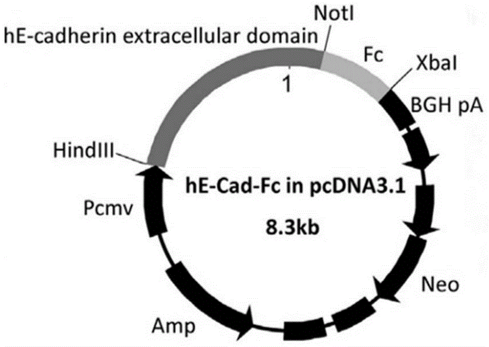
[0057] Embodiment 1 obtains pcDNA3.1-hE-cad-Fc gene plasmid

[0058] Human epithelial cell adhesion factor (hE-cadherin) ectodomain (hE-cad) and human immunoglobulin (IgG 1) Fc fragment (Fc) were fused by gene recombination technology to obtain a plasmid containing the target gene hE-cad and Fc pcDNA3.1-hE-cad-Fc. That is: using the full length of human E-cadherin mRNA (Gene bank: NM 004360.3) as a template, the synthetic primers were used to specifically amplify the hE-cadherin ectodomain fragment. The primer sequences are:

[0059] 5'-CGCAAGCTTATGGGCCCTTG-GAGCCGCAGC-3';

[0060] 5'-TTGCGGCCGCAGGCAGGAATTTGCAATCCTGC-3'.

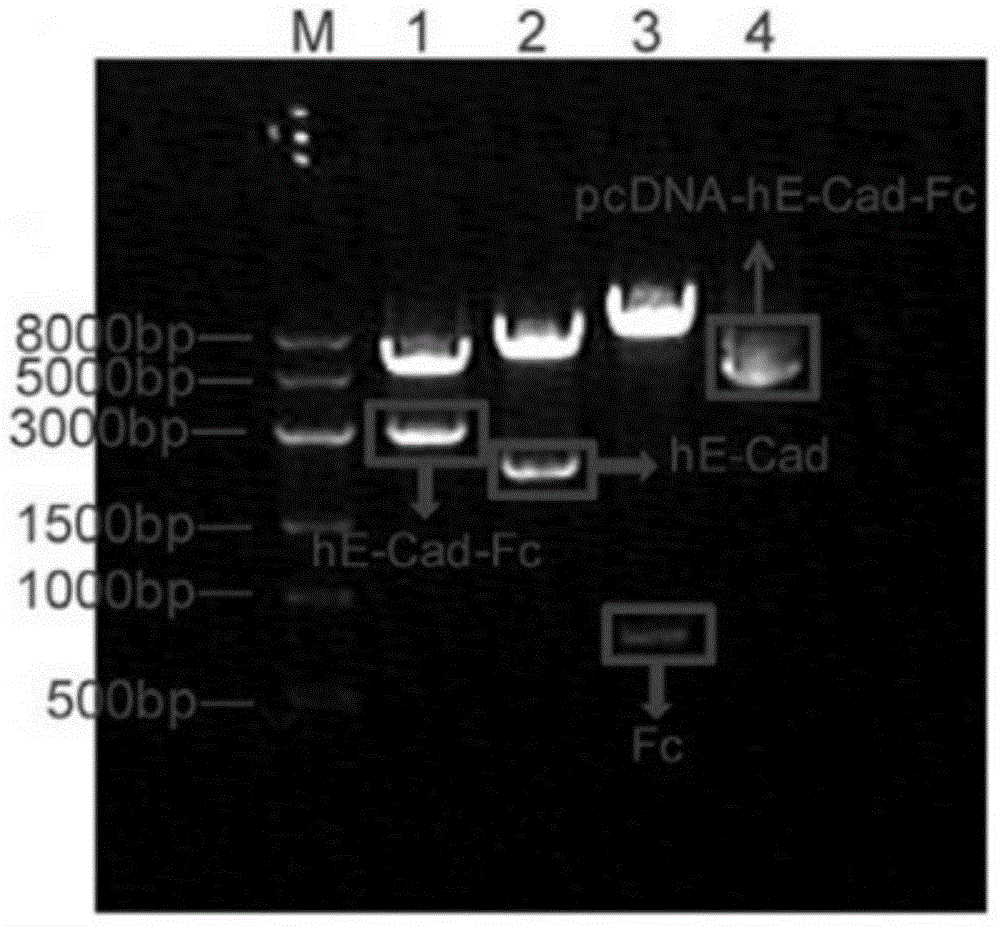
[0061] like figure 1 As shown, the PCR product was analyzed by 1% agarose gel electrophoresis, and the gel-recovered PCR product was double-digested with Hind III and Not I, and inserted between the Hind III and Not I sites of pcDNA3.1-Fc. Plasmid pcDNA3.1-Fc was donated by Professor Toshihiro Akaike of Tokyo Institute of Technology.

[0062] like fig...

Embodiment 2

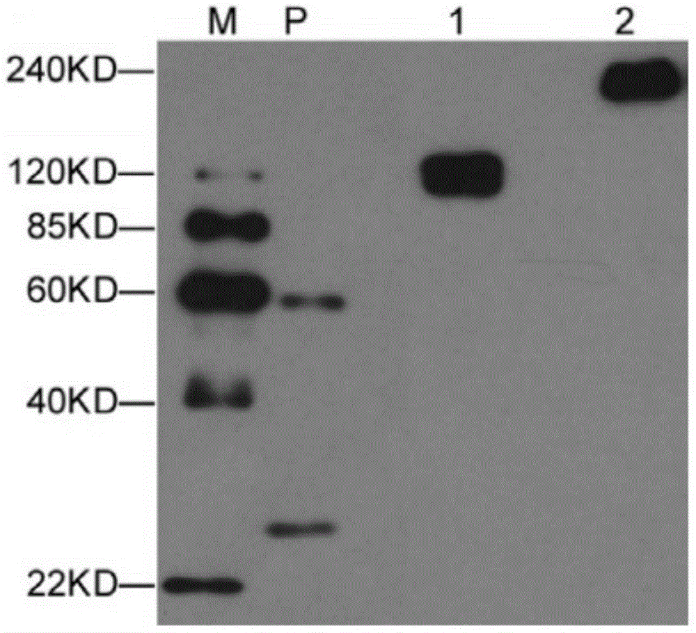
[0064] Embodiment 2 obtains hE-cad-Fc fusion protein

[0065] The plasmid pcDNA3.1-hE-cad-Fc containing the target gene constructed in Example 1 was transfected into 293F cells, cultured in suspension for 72 hours, the cell culture supernatant was collected, and the hE-cad-Fc fusion protein was purified by rProtein AFF column . The purified fusion protein was detected by immunoblotting using an anti-E-cadherin antibody, as image 3 As shown, there are no other non-specific exposure bands in the figure, and the band with a molecular weight of about 120KD is reduced E-cadherin ( image 3 Middle 1), the band at 240KD is non-reduced E-cadherin ( image 3 Medium 2). This result shows that the hE-cad-Fc fusion protein prepared by the present invention has a base sequence as shown in 1 in the sequence table, and the fusion protein can form a stable dimer structure in a non-reduced state.

Embodiment 3

[0066] Example 3 Preparation of hE-cad-Fc fusion protein matrix-modified surface-modified PLGA microspheres

[0067] First, microspheres with a particle size range of 15±5 μm were prepared by chemically synthesizing the polymer PLGA and placed in an EP tube; then the above hE-cad-Fc fusion protein solution was diluted to 10 μg / ml, soaked into the microspheres, and incubated at 37°C for 2 hours; the supernatant was discarded by centrifugation, rinsed with PBS three times to remove the unimmobilized fusion protein hE-cad-Fc, and obtained hE-cad-Fc matrix-modified PLGA microspheres, which were named hE-cad- Fc-PLGA.

[0068] The invention detects the effect of the protein concentration of the hE-cad-Fc solution on the surface modification of the matrix of PLGA microspheres by an Elisa method. The hE-cad-Fc substrate method is the same as above. Namely: after obtaining hE-cad-Fc-PLGA, add 1% BSA to block at room temperature for 1 hour, add anti-E-cadherin antibody and incubate a...


PUM

PropertyMeasurementUnit
particle diameteraaaaaaaaaa

Abstract

The invention provides a three-dimensional complex cell aggregate model as well as a preparation method and application thereof. According to the model, a natural or chemically synthesized polymer is used as a matrix material to prepare submicron-scale microspheres, and cell factors are embedded in or fixed on the surface of the submicron-scale microspheres and are co-cultured with cells to form a complex cell aggregate containing the microspheres, so that the biological activity of the cells in the aggregate can be regulated and controlled from inside to outside. The model can be used for researches of propagation and differentiation of stem cells in a three-dimensional state, overcomes the problems that the cell aggregate is heterogeneous in composition/structure, the cell proliferation efficiency is low and the directional induced differentiation efficiency is low due to non-uniform mass transfer and short acting time of liquid factors in a culture medium during the traditional three-dimensional culture of the stem cells, can be used for stimulating interaction between cells and cells, cells and an extracellular matrix as well as cells and soluble factors, thereby providing a theoretical and technical support for in vitro construction and optimization of a stem cell culture technology, and tissue engineering construction of various tissue and organ analogs or equivalents.

Description

technical field [0001] The invention relates to the technical field of in vitro cell culture, in particular to a three-dimensional composite cell aggregate model and its preparation method and application. Background technique [0002] In the body, cells are in a highly informational microenvironment, which includes various physical and chemical signals that change dynamically in time / space, and cells are regulated by various signal stimuli to achieve their specific biological functions. This interaction between cells and the microenvironment includes a variety of complex biochemical, biomechanical, and bioelectrical signal stimuli responses from surrounding cells, extracellular matrix, and soluble factors. Among them, the interaction between cells occurs through direct contact or paracrine soluble factors. This intercellular communication is an important link in maintaining the structure and function of cells, tissues and organs, and is also a key factor in regulating tiss...

Claims


Application Information

Patent Timeline
Patent Type & Authority Applications(China)
IPC IPC(8): C12N5/0735C12N5/0775
Inventor 杨军张妍李素华徐建斌
Owner NANKAI UNIV
Why Eureka
Try Eureka