Preparation method for nanofiber porous scaffold having compression elasticity in wet state

A nanofiber and compressive elasticity technology is applied in the field of preparation of three-dimensional porous scaffolds, which can solve the problems of poor mechanical properties of scaffolds, poor mechanical properties of three-dimensional scaffolds, and difficulty in application, and achieve the effects of high compressive elasticity and excellent water absorption performance.

Active Publication Date: 2015-12-02
诺一迈尔(苏州)生命科技有限公司
View PDF3 Cites 38 Cited by
  • Summary
  • Abstract
  • Description
  • Claims
  • Application Information

AI Technical Summary

Problems solved by technology

Electrospinning technology is a simple and rapid method for preparing nanofibers, but the scaffolds prepared by this technology are generally fibrous membranes composed of nanofiber accumulations, and the prepared three-dimensional scaffolds have poor mechanical properties and are difficult to achieve real practical applications (B.Sun , Y. Long, H. Zhang, M. Li, J. Duvail, X. Jiang and H. Yin, Advances in three-dimensional nanofibrous macrostructures via electrospinning, Progress in Polymer Science, 39 (2014) 862-890.)
The limitation of self-assembly technology is that i

Method used


Image

  • Preparation method for nanofiber porous scaffold having compression elasticity in wet state
  • Preparation method for nanofiber porous scaffold having compression elasticity in wet state
  • Preparation method for nanofiber porous scaffold having compression elasticity in wet state

Examples


Embodiment 1

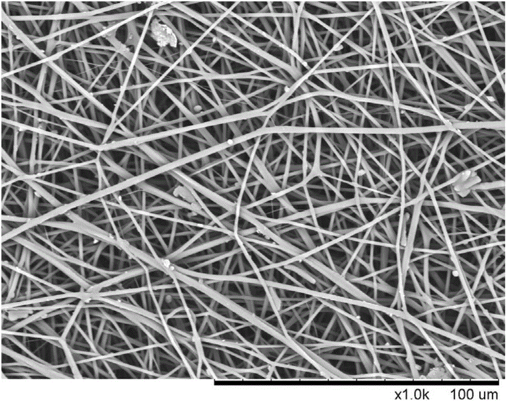
[0027] Gelatin and polylactic acid (mass ratio 1:5) were dissolved in hexafluoroisopropanol, with a total mass fraction of 11%. Inhale the solution into the syringe, control the advance rate of the micro-injection pump to 5mL / h, connect a 15KV high-voltage power supply to the syringe needle, and receive it with aluminum foil at a receiving distance of 15cm. Gelatin / PLA nanofiber membranes were obtained by electrospinning.

Embodiment 2

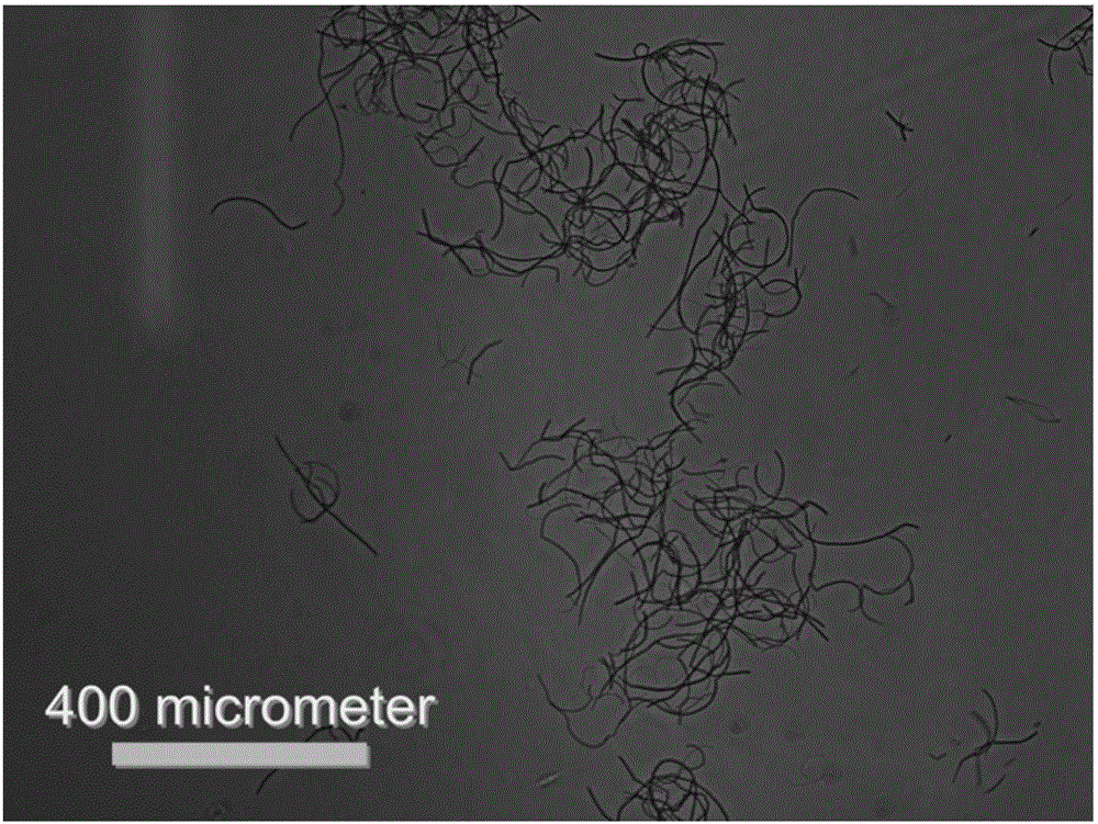
[0029] Gelatin and polylactic acid (mass ratio 1:5) were dissolved in hexafluoroisopropanol, with a total mass fraction of 11%. Inhale the solution into the syringe, control the advance rate of the micro-injection pump to 5mL / h, connect a 15KV high-voltage power supply to the syringe needle, and receive it with aluminum foil at a receiving distance of 15cm. Gelatin / PLA nanofiber membranes were obtained by electrospinning. Cut the nanofibrous membrane into a sheet with a size of about 0.5×0.5 cm with scissors. Then weigh 1 g of the nanofiber membrane and add it to a beaker containing 100 mL of tert-butanol, and use a high-speed pulverizer to pulverize the nanofiber membrane into a uniform short fiber solution.

Embodiment 3

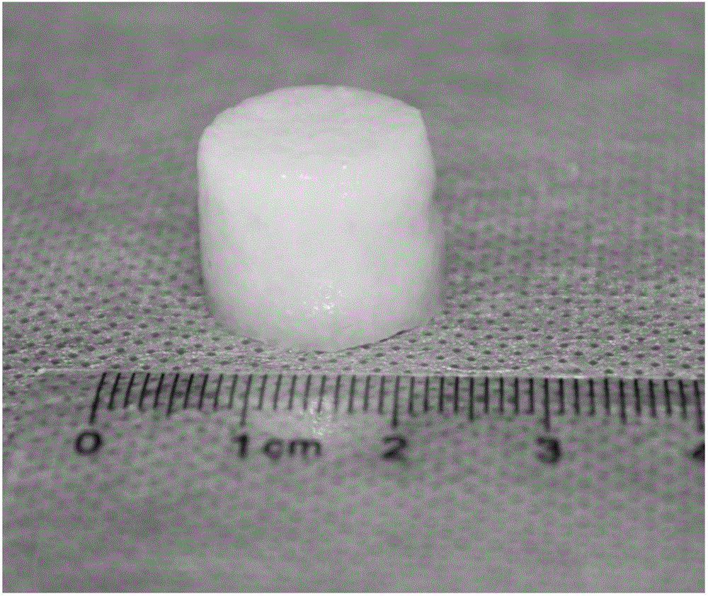
[0031] Gelatin and polylactic acid (mass ratio 1:5) were dissolved in hexafluoroisopropanol, with a total mass fraction of 11%. Inhale the solution into the syringe, control the advance rate of the micro-injection pump to 5mL / h, connect a 15KV high-voltage power supply to the syringe needle, and receive it with aluminum foil at a receiving distance of 15cm. Gelatin / PLA nanofiber membranes were obtained by electrospinning. Cut the nanofibrous membrane into a sheet with a size of about 0.5×0.5 cm with scissors. Then weigh 1 g of the nanofiber membrane and add it to a beaker containing 100 mL of tert-butanol, and use a high-speed pulverizer to pulverize the nanofiber membrane into a uniform short fiber solution. Then, add the short fiber solution into the mold, put it in a refrigerator at -80° C., and freeze it for 1 hour. Finally, put it into a vacuum freeze dryer and dry it for 24 hours to obtain a three-dimensional scaffold. Put the scaffold into a mixed solution of glutaral...


PUM


Abstract

The invention relates to a preparation method for a nanofiber porous scaffold having compression elasticity in a wet state. The preparation method comprises the following steps: dissolving gelatin and polylactic acid in a solvent so as to obtain a mixed solution, and carrying out electrostatic spinning so as to obtain a nanofiber membrane; feeding the nanofiber membrane into tertiary butanol, smashing, freezing and freeze-drying so as to obtain a non-crosslinked freeze-dried scaffold; immersing the non-crosslinked freeze-dried scaffold into cross-linking liquid, washing, soaking in deionized water, freezing and freeze-drying so as to obtain the nanofiber porous scaffold. The nanofiber porous scaffold prepared through the preparation method has degradability, biocompatibility and relatively high porosity, and has a certain compression resistance at the wet state, can quickly stop bleeding, is beneficial to cell adhesion, cell proliferation and tissue regeneration, and can be applicable to hemostatic materials, cartilage tissue engineering, skin tissue engineering and other fields.

Description

technical field [0001] The invention belongs to the field of preparation of three-dimensional porous supports, in particular to a method for preparing nanofiber porous supports with compressive elasticity in a wet state. Background technique [0002] Cells, scaffolds and growth factors are the three major elements of tissue engineering. In the process of using tissue engineering methods to repair damaged tissues, the preparation and selection of scaffolds is an important link. Excellent tissue engineering scaffolds should simulate natural tissues and organs in terms of structure and physiological functions, provide mechanical support and three-dimensional space for cell growth, and meet the requirements of cell adhesion, proliferation, migration and differentiation (S.Yang, K.-F . Leong, Z. Du and C.-K. Chua, The design of scaffolds for use in issue engineering. Part I. Traditional factors, Tissue engineering, 7 (2001) 679-689). Therefore, the preparation of three-dimensio...

Claims


Application Information

Patent Timeline
IPC IPC(8): A61L27/26A61L27/52A61L27/50
Inventor 莫秀梅陈维明
Owner 诺一迈尔(苏州)生命科技有限公司
Why Eureka
Try Eureka