
Primary isolation method of animal skin fibroblasts

A fibroblast and animal skin technology, applied in the field of stem cell culture, can solve the problems of cell mechanical damage, low attachment stability, and low cell viability, so as to improve the separation efficiency and quality, enhance the attachment efficiency, and increase the number of cells Quantity effect

Inactive Publication Date: 2016-03-30
GUANGZHOU SALIAI STEMCELL SCI & TECH CO LTD
View PDF1 Cites 3 Cited by
  • Summary
  • Abstract
  • Description
  • Claims
  • Application Information

AI Technical Summary

Problems solved by technology

[0004] For the enzymatic digestion method, although the cells can be obtained in a short period of time, the enzyme itself has a certain influence on the adhesion of the cells, and repeated centrifugation also causes certain mechanical damage to the cells, resulting in low cell viability and poor adhesion rate. Low
Compared with the enzymatic hydrolysis method, the tissue block attachment method has simpler steps, but the cycle is long, the attachment stability is low, and the cell crawling rate is low.

Method used


Image

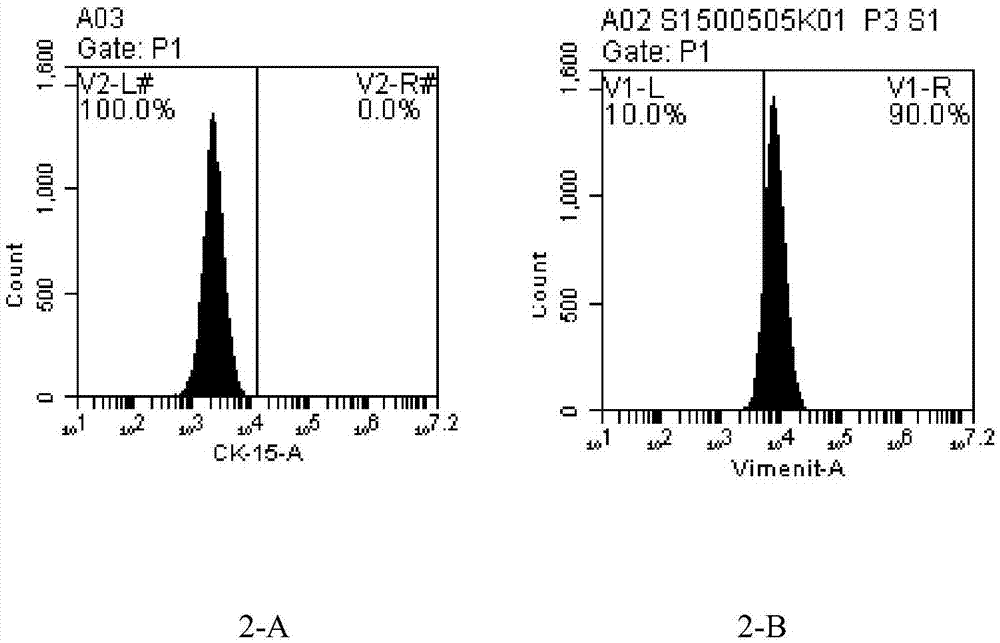
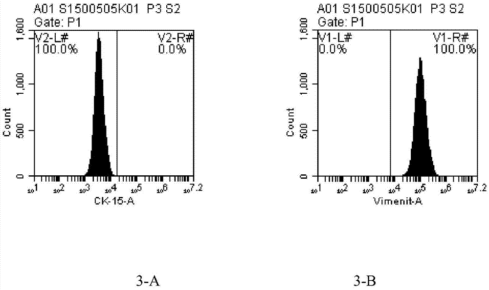
  • Primary isolation method of animal skin fibroblasts
  • Primary isolation method of animal skin fibroblasts
  • Primary isolation method of animal skin fibroblasts

Examples


Embodiment 1

[0020] A kind of mouse skin fibroblast primary separation method of the present embodiment, it comprises the steps:

[0021] 1.1 Select the abdominal skin of the mouse, use curved scissors to slightly cut off the hair, disinfect the skin with iodine and alcohol, make a circular full-thickness skin wound with a diameter of 2.0 cm, expose the wound and continue to raise.

[0022] 1.2 After skin injury (3-5) days, the mice were sacrificed, the injured skin area was removed, the subcutaneous tissue and excess blood vessels were removed, and the skin was washed 3 times with PBS containing 2% double antibody.

[0023] 1.3 Cut the skin to 0.5mm with ophthalmic scissors 3 size organization block.

[0024] 1.4 Pour in an appropriate amount of PBS containing 1% double antibody, centrifuge at a low speed of 1000rpm / min for 5min, remove floating tissue pieces, and discard the supernatant.

[0025] 1.5 Add 0.2% gelatin 1mL / dish to a 9cm cell culture dish in advance and incubate for 30min...

Embodiment 2

[0028] This embodiment is the primary isolation method of conventional conventional mouse skin fibroblasts, comprising the following steps:

[0029] 2.1 The mice were killed, the hair was slightly trimmed with curved scissors, the skin was disinfected with iodine and alcohol, the skin tissue with a diameter of 2 cm was removed, and the skin was washed 3 times with PBS containing 2% double antibody.

[0030] 2.2 Cut the skin to 0.5mm with ophthalmic scissors 3 size organization block.

[0031] 2.3 Pour in an appropriate amount of PBS containing 1% double antibody, centrifuge at a low speed of 1000rpm / min for 5min, remove residual trypsin floating tissue pieces and pour off the supernatant.

[0032] 2.4 The centrifuged tissue pieces were evenly inoculated in a 9cm cell culture dish, and 2ml of medium (DMEM+10%FBS+2% double antibody) cell culture solution was added, and placed at 37°C, 5%CO 2 cultured in an incubator.

Embodiment 3

[0034] This embodiment is a comparison of the cell growth status after the separation of mouse skin fibroblasts

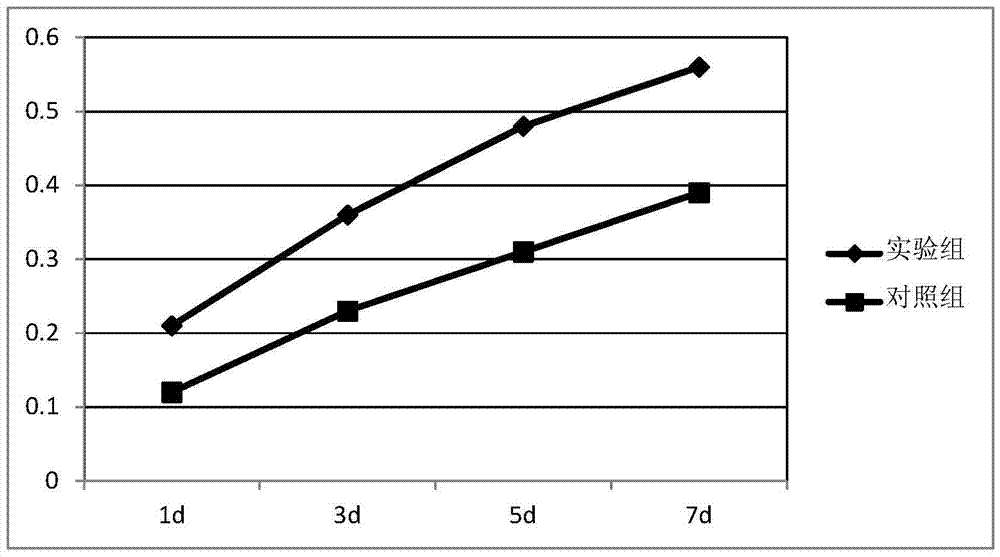
[0035] Three different batches of mice were treated with the methods of Example 1 (experimental group) and Example 2 (control group), respectively, and the cell growth of the tissue block was observed every day after inoculation.

[0036] Table 1: Comparison of the number of cells in the experimental group and the control group

[0037]

[0038] As can be seen from Table 1, the number of cells separated by the experimental group is more than 2 times that of the control group, which proves that the scheme of the present invention can greatly improve the number of cells separated.


PUM

PropertyMeasurementUnit
diameteraaaaaaaaaa

Abstract

The invention discloses a primary isolation method of animal skin fibroblasts. Disinfected animal skin is selected, and a full-layer skin wound is formed; an injured skin area is taken down after the skin is injured within 3-5 days, subcutaneous tissue and extra blood vessels are removed completely, and the skin is cleaned with PBS (phosphate buffer saline) containing 2% of a penicillin-streptomycin solution for 2-3 times; the skin is cut into tissue pieces ranging from 0.1 mm<3> to 0.5 mm<3> in size; PBS containing 1% of the penicillin-streptomycin solution is poured, centrifugation is performed at the low speed of 1,000 rpm / min for 5 min, and residual floating tissue pieces and a residual supernatant are removed; a cell culture dish covered with 0.2% of gelatin is evenly inoculated with centrifuged tissue pieces, a cell culture fluid containing 2 ml of a DMEM (Dulbecco modified eagle medium), 10% of the FBS and 2% of the penicillin-streptomycin solution is added, and the culture dish is placed in a 5% CO2 incubator at the temperature of 37 DEG C for culture. A skin injury healing mechanism is used, a proper healing period is selected, a large number of proliferative fibroblasts are obtained, meanwhile, removal of the subcutaneous tissue facilitates climbing out of the fibroblasts of corium layers, the number of cells can be increased, the cells can climb out more easily, the cell climbing-out cycle is shortened, and the adherence efficiency is enhanced.

Description

technical field [0001] The invention relates to a method for primary separation of cells, in particular to a method for primary separation of animal skin fibroblasts, which belongs to the field of stem cell culture. Background technique [0002] At present, fibroblasts are mainly used to study the aging of cells, the damage of various external factors to cells, the malignant transformation of cells in vitro, and some congenital metabolic abnormalities and enzyme defects. Because skin fibroblasts are easy to obtain and grow in vitro, the culture of skin fibroblasts has been widely used in basic and clinical medical research. [0003] For the acquisition of fibroblasts, there are mainly two primary separation methods currently available: (1) Enzyme digestion. Use trypsin or collagenase to digest the skin tissue rinsed with double-antibody-containing PBS and shred until the cells are separated, pass through 200 sieves, centrifuge, resuspend, count the cell density, and inocula...

Claims


Application Information

Patent Timeline
Patent Type & Authority Applications(China)
IPC IPC(8): C12N5/071
Inventor 葛啸虎陈海佳王一飞吴子杰李平
Owner GUANGZHOU SALIAI STEMCELL SCI & TECH CO LTD
Learn More