Stem cell culture medium and method for culturing endometrium stem cells
An endometrial stem cell and cell culture technology, applied in the field of stem cell culture medium, can solve the problems of high price, easy aging of cells, and many medium components, and achieve low cost, maintain activity and stemness, and fast cell proliferation efficiency. Effect
- Summary
- Abstract
- Description
- Claims
- Application Information
AI Technical Summary
Problems solved by technology
Method used
Image
Examples
Embodiment 1
[0034] Stem cell medium, according to the volume content of the total volume percentage, low-sugar DMEM medium accounted for 82%, fetal bovine serum accounted for 15%, penicillin and streptomycin double antibody solution accounted for 1%; gentamicin sulfate accounted for 1%; glutamine Amide accounts for 1%.
Embodiment 2
[0036] The method for cultivating endometrial stem cells using the stem cell medium provided in Example 1 comprises the following steps:
[0037] Step 1: Use the menstrual blood collection device to collect menstrual blood and endometrial tissue, and transfer the menstrual blood to a sterile centrifuge tube containing sodium heparin, transfer the endometrial tissue to a sterile culture dish, and use a sealing film to separate the centrifuge tube and culture Seal the opening of the dish, and transport it to the laboratory at low temperature (4-8°C) within 24 hours;
[0038] Step 2: Fully mix the menstrual blood in the centrifuge tube described in step 1 with an equal volume of normal saline, which contains 1% penicillin and streptomycin double antibody solution for inhibiting bacterial growth. Mix menstrual blood and normal saline evenly, superimpose with Ficoll separation solution 1:1, centrifuge at 800g for 20min, and collect buffy coat mononuclear cells. Then resuspend the ...
Embodiment 2
[0043] The endometrial stem cell identification method described in Example 2 is as follows:
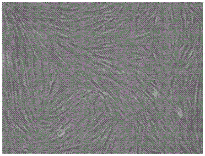
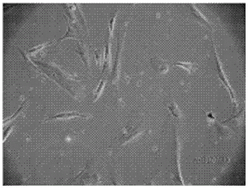
[0044] 1) Morphological characteristics: Place the cell culture flask under an inverted microscope, connect the microscope to a camera device, adjust the field of view of the cells and take pictures. The cells are in the shape of long spindles and arranged in clusters.
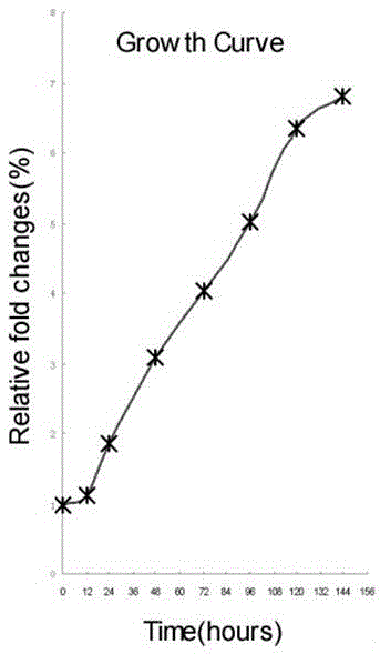
[0045] 2) Draw the cell growth curve: resuspend the cells after subculture to obtain a cell suspension, and add the suspension evenly to a 1-well plate; start counting the cells at 24 hours, count once every 12 hours thereafter, and take 3 wells of cells each time, Count them separately, and take the average value of 3 wells for the counting results, and count continuously for 6 days. According to the cell counting results, the growth curve was drawn with unit cell number (cell number / ml) as the ordinate and time as the abscissa.
[0046] 3) Detection of cell surface markers: Directly label cell surface molecules wi...
PUM
 Abstract
Description
Claims
Application Information
 - R&D
- Intellectual Property
- Life Sciences
- Materials
- Tech Scout
- Unparalleled Data Quality
- Higher Quality Content
- 60% Fewer Hallucinations
Browse by: Latest US Patents, China's latest patents, Technical Efficacy Thesaurus, Application Domain, Technology Topic, Popular Technical Reports.
© 2025 PatSnap. All rights reserved.Legal|Privacy policy|Modern Slavery Act Transparency Statement|Sitemap|About US| Contact US: help@patsnap.com


