Tumor-targeted diagnosis and treatment integrated fluorescent probe

A tumor-targeting and probe technology, applied in the field of fluorescent probes for tumor-targeted diagnosis and treatment, can solve problems such as inability to feedback and monitor, and achieve the effects of improving solubility, simple preparation method, and simple purification process

Active Publication Date: 2016-06-22
WUHAN UNIV
View PDF5 Cites 12 Cited by
  • Summary
  • Abstract
  • Description
  • Claims
  • Application Information

AI Technical Summary

Problems solved by technology

In addition, individual differences and the complexity of the physiological environment may also cause false positive signals
Therefore, re

Method used


Image

  • Tumor-targeted diagnosis and treatment integrated fluorescent probe
  • Tumor-targeted diagnosis and treatment integrated fluorescent probe
  • Tumor-targeted diagnosis and treatment integrated fluorescent probe

Examples


Embodiment 1

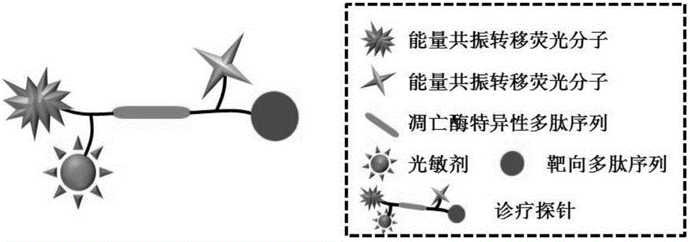
[0048] [Example 1] Synthesis of fluorescent probes for tumor targeting diagnosis and treatment

[0049] Protoporphyrin-Lysine (Fluorescein)-Serine-Aspartic Acid-Glutamic Acid-Valine-Aspartic Acid-Serine-Lys (Dimethylaminoazobenzene)-Arginine- Synthesis of Glycine-Aspartic Acid, PpIX-K(FAM)SDEVDSK(Dabcyl)RGD) at room temperature:

[0050] (1) Add 0.5g of ammonia resin (0.525mmol / g) to the reactor containing 10mL of re-steamed N,N-dimethylformamide, and wait until the ammonia resin is in N,N-dimethylformamide After swelling for 2h at room temperature, N,N-dimethylformamide was removed.

[0051] (2) Add 10 mL of 20% (V / V) piperidine / N,N-dimethylformamide (that is, the volume ratio of piperidine to N,N-dimethylformamide is 2:8) into the reactor. After reacting at room temperature for 15 minutes, remove the solvent; repeat the reaction by adding piperidine / N,N-dimethylformamide solution to cut off the FMOC protecting group. After the reaction, remove the solvent and use N,N-dimethylform...

Embodiment 2

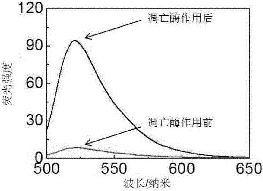
[0063] [Example 2] Detection of the response of a fluorescent probe for tumor targeting diagnosis and treatment to apoptotic enzyme-3

[0064] Dissolve the probe in the HEPES buffer solution and configure it as a working solution of 1 μmol / L. The apoptotic enzyme-3 (1U) was added to the buffer solution containing the probe, and the working solution was diluted with the buffer solution to a final concentration of 0.5 micromol / L. Fluorescence spectrometer (LS55 fluorescence spectrophotometer, Perkin-Elmer) was used to detect the fluorescence intensity of luciferin in the solution immediately after the apoptotic enzyme-3 was added and 11 hours after the apoptotic enzyme-3 was added. The excitation wavelength of fluorescein is: 465 nm.

[0065] The result is figure 2 As shown, the luciferin intensity of the probe in the solution was weak when the apoptotic enzyme-3 was just added, and after 11 hours of interaction with the apoptotic enzyme-3, the luciferin intensity of the probe was ...

Embodiment 3

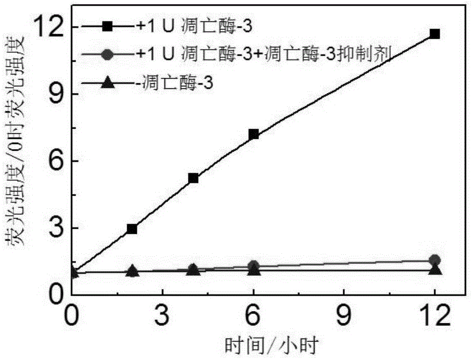
[0066] [Example 3] Specific detection of the response of apoptotic enzyme-3 by fluorescent probes for tumor targeting diagnosis and treatment

[0067] The apoptotic enzyme-3 (1U) was incubated with a commercial apoptotic enzyme-3 specific inhibitor (Ac-DEVD-CHO, 50 μmol / L) at 37 degrees Celsius for 2 hours. The probe solution was prepared into a working solution of 1 micromol / L in the HEPES buffer solution. Add apoptotic enzyme-3 (1U) and apoptotic enzyme-3 (1U) incubated with the apoptotic enzyme inhibitor to the buffer solution containing the probe, and dilute the probe concentration with the buffer solution to a final concentration of 0.5 μmol / Rise. Use a fluorescence spectrometer to record how the fluorescence of the probe solution without apoptotic enzyme-3, apoptotic enzyme-3, and apoptotic enzyme-3 and inhibitors changes over time. The excitation wavelength of fluorescein: 465 nm; the emission wavelength of collected fluorescein: 520 nm.

[0068] The result is image 3 A...


PUM

PropertyMeasurementUnit
Excitation wavelengthaaaaaaaaaa

Abstract

The invention discloses a tumor-targeted diagnosis and treatment integrated fluorescent probe which takes a tumor-targeted polypeptide sequence and an apoptosis enzyme specific recognition polypeptide sequence as a framework and comprises a fluorescence quenching molecular fluorescence pair and a photodynamic treatment photosensitizer. The diagnosis and treatment integrated fluorescent probe can achieve targeted treatment of tumors, and reduce toxic and side effects of the photodynamic treatment photosensitizer; a treatment result can be assessed in situ accurately in real time while the tumors are subjected to photodynamic treatment. The tumor-targeted diagnosis and treatment integrated fluorescent probe can also be used as a common fluorescent probe and used for screening of tumor treatment medicines and cell apoptosis imaging in a proportional fluorescence imaging manner. The tumor-targeted diagnosis and treatment integrated fluorescent probe can realize early detection of tumor treatment feedbacks and has great significance in promotion of accurate treatment of tumors and personal treatment.

Description

Technical field [0001] The invention relates to a fluorescent probe for detecting tumor markers and combining with photodynamic therapy to achieve targeted therapy, and a preparation method and application thereof. Background technique [0002] Cancer is a serious threat to human health. Chemotherapy, radiotherapy and gene therapy have been widely used in cancer treatment research, but they are facing the problem of how to further improve the effect of cancer treatment and reduce the toxic and side effects of drugs. Realizing targeted drug delivery to tumor sites has become today's clinical medicine and biomedicine One of the research hotspots in the field; the emergence of multidrug resistance, changes in the tumor microenvironment, and individual differences lead to the failure of tumor treatment, realize personalized treatment methods for tumor treatment, and timely feedback the tumor treatment effect, quickly Optimizing the treatment plan will more effectively solve the prob...

Claims


Application Information

Patent Timeline
IPC IPC(8): C09K11/06C07K7/06A61K41/00G01N21/64
CPCA61K41/0061A61K41/0071C07K7/06C09K11/06C09K2211/1074C09K2211/1088G01N21/6486G01N2021/6417
Inventor 张先正李仕颖成红曾旋冯俊
Owner WUHAN UNIV
Why Eureka
Try Eureka