Preparation and application of magnetic resonance and near-infrared fluorescence targeted nanoprobes
A nano-probe and near-infrared technology, which is applied in the field of nuclear magnetic and fluorescence imaging nanomaterials, can solve the problems of probe tumor targeting and multi-modal multifunctional performance, so as to enhance MRI imaging ability and prolong the effective time , the effect of extending the window time
- Summary
- Abstract
- Description
- Claims
- Application Information
AI Technical Summary
Problems solved by technology
Method used
Image
Examples
Embodiment 1
[0032] MR&NIR-NPs preparation method:
[0033] (1) Synthesis of PEI-PEG-FA-DOTA-Gd carrier material
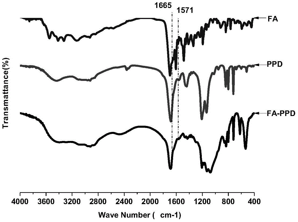
[0034] The EDC activated ester of PEG-COOH reacts covalently with PEI in a certain ratio (m / m=1:1) to prepare PEI-PEG, and then PEI-PEG is covalently grafted with TB-DOTA and FA through carboxyl esterification ( PEI-PEG:TB-DOTA:FA(m / m / m)=300:53.2:20.6), prepare PEI-PEG-FA-DOTA-TB, TFA / DMF deprotection, then chelate gadolinium acetate (feeding amount is Add 15.05 mg of gadolinium acetate per 100 mg of PEI-PEG-FA-DOTA-TB) to prepare PEI-PEG-FA-DOTA-Gd, and the infrared spectrum of the material is as follows figure 1 .
[0035] (2) Preparation of MR&NIR-NPs nanoparticles
[0036] Dissolve material PEI-PEG-FA-DOTA-Gd in deionized water or PBS (PH=7.4), concentration 2mg / ml, ultrasonic 100W ultrasonic 10s / pause 5s / 10 times, prepare PEI-PEG-FA-DOTA-Gd solution. The material ICG was dissolved in deionized water or PBS (PH=7.4) at a concentration of 1 mg / ml to prepare an ICG solu...
Embodiment 2
[0038] Characterization of MR&NIR-NPs:
[0039] (1) Morphology and size of MR&NIR-NPs
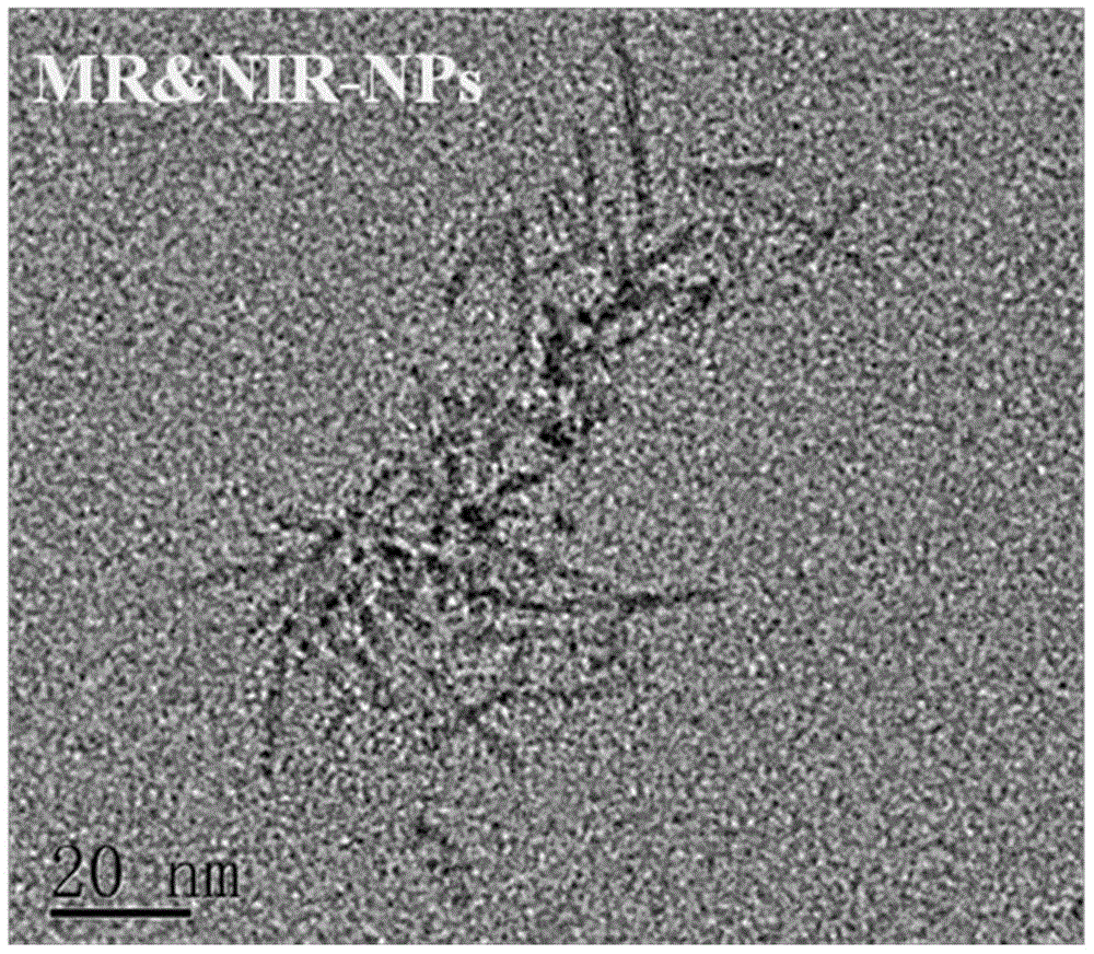
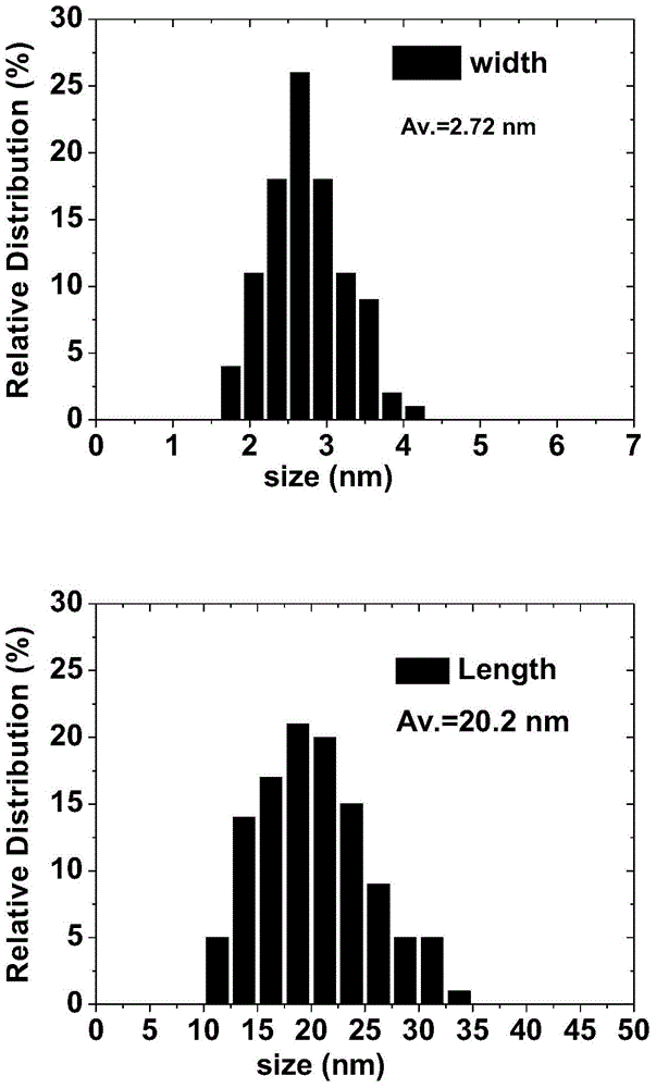
[0040] figure 2 It is a high-resolution transmission electron micrograph of MR&NIR-NPs, which shows that MR&NIR-NPs form a fibrous nanostructure with a narrow size distribution. image 3 It is a statistical analysis of the size distribution of electron microscope results. The results show that the average fiber diameter is 2.72nm, the average fiber length is 20.2nm, and the distribution is uniform.
[0041] (2) MR imaging performance evaluation of MR&NIR-NPs
[0042] Figure 4 is the MR imaging performance evaluation of MR&NIR-NPs. In the figure, the concentrations of MR & NIR-NPs are 1mg / ml, 0.5mg / ml, 0.25mg / ml, 0.125mg / ml, 0.0625mg / ml, 0mg / ml (the probes prepared in Example 1 were diluted with ultrapure water Obtain) MR imaging, imaging conditions: Tr=300ms, Te=19ms. The results show that the MR&NIR-NPs probe has good MR imaging ability.
[0043] (3) Evaluation of near-infrared fluo...
Embodiment 3
[0048] In vivo tumor targeted imaging of MR&NIR-NPs:
[0049] Figure 7 For MR&NIR-NPs targeted tumor MRI imaging results, Figure 8 It is the near-infrared fluorescence imaging results of MR&NIR-NPs targeting tumors. Imaging method: 7.5 mg / ml of MR&NIR-NPs solution and 7.5 mg / ml of control group (PPD-ICG) solution were prepared with normal saline as a solvent, and 100 μl per 10 g of nude mice body weight were injected into U87 glioma-bearing tumors through the tail vein in nude mice. At time points 0 (no injection), 24, 48, 72, and 96 h, MR imaging was performed under the conditions of Tr=300 ms, Te=19 ms, Av=2 times, slice thickness 2.5 mm, and nude mice were anesthetized during the process. . At the time points of 25min, 65min, 48h, 72h, and 96h, NIR imaging was performed under the conditions of 735nm excitation and 780nm-950nm acquisition, and nude mice were anesthetized during the process. The results show that MR&NIR-NPs have remarkable NMR and NIR targeted imaging ...
PUM
| Property | Measurement | Unit |
|---|---|---|
| Diameter | aaaaa | aaaaa |
Abstract
Description
Claims
Application Information
 - R&D
- Intellectual Property
- Life Sciences
- Materials
- Tech Scout
- Unparalleled Data Quality
- Higher Quality Content
- 60% Fewer Hallucinations
Browse by: Latest US Patents, China's latest patents, Technical Efficacy Thesaurus, Application Domain, Technology Topic, Popular Technical Reports.
© 2025 PatSnap. All rights reserved.Legal|Privacy policy|Modern Slavery Act Transparency Statement|Sitemap|About US| Contact US: help@patsnap.com


