Self-assembly collagen template tissue engineering material as well as preparation method and application thereof

A technology of tissue engineering and self-assembly, applied in medical science, prosthesis, etc., can solve the problems of complex preparation, cytotoxic side effects, fast degradation, etc., and achieve the effect of simple preparation method, controllable process parameters, and good biological activity

Active Publication Date: 2016-07-27
CHENGDU UNIV
View PDF6 Cites 4 Cited by
  • Summary
  • Abstract
  • Description
  • Claims
  • Application Information

AI Technical Summary

Problems solved by technology

However, this method is complicated to prepare, and the photocrosslinking time is long, which leads to the sinking of loaded cells, poor mechanical properties, and too fast degradation, which cannot match the dynamic growth of cells; at the same time, the crosslinking agent used, methacrylic anhydride and photoinitiators, which have certain toxic side effects on cells

Method used


Image

  • Self-assembly collagen template tissue engineering material as well as preparation method and application thereof
  • Self-assembly collagen template tissue engineering material as well as preparation method and application thereof
  • Self-assembly collagen template tissue engineering material as well as preparation method and application thereof

Examples


Embodiment 1

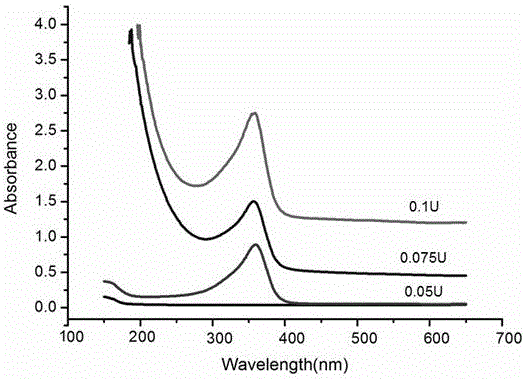
[0058] Dissolve 10g of collagen in 100ml of Tris buffer solution, stir magnetically at 35~45°C until clarified, take the glycerol solution and add it to the collagen solution in a ratio of (10:1) and mix evenly; Aminamidase is dissolved in deionized water, the amount added varies with the enzyme activity, and the enzyme activity is between 0.025-0.1. Mix the two prepared solutions at 35-45°C to make them cross-linked and gel The coagulation time of the body varies with the amount of enzyme added.

[0059] Table 1: Clotting time corresponding to different enzyme additions

[0060]

[0061] Through the ultraviolet spectrometer, the amino cross-linking peak appears near 350nm, the degree of cross-linking gradually increases with the addition of enzyme, the absorption peak is enhanced, and the curing time is shortened, but the curing time at a higher temperature (36.5°C) is shorter than that at room temperature ( 25°C) the curing time is slightly longer, indicating that the cr...

Embodiment 2

[0063] Dissolve 2g of collagen in 20ml of Tris buffer solution, stir magnetically at 35~45°C until clarified, take glycerol solution and add it to the collagen solution in a ratio of (10:1) and mix evenly; mix 0.5g of bio-based Transglutaminase was dissolved in 1ml of deionized water, the two solutions were magnetically stirred and mixed evenly, poured onto a glass plate covered with polyethylene film, and dried at 25-37°C for 24 hours to obtain a 2D collagen film.

Embodiment 3

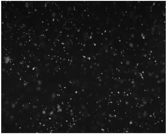
[0065] Cell experiment: Mix the collagen / glycerol solution prepared in Example 1 with the cultured human umbilical vein endothelial cells (HUVECs) cells within 10 minutes, then add 0.075U transaminase, and perform 3D in a transwell culture plate Cultivate, observe its cell growth status under an optical microscope, and evaluate its cell proliferation and life and death status.

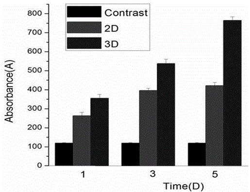
[0066] The result is as figure 1 As shown, the cells are evenly distributed in the collagen gel. After 24 hours of culture, it is found that there are red blood cells, that is, dead cells. During the daily replacement of the cell fluid, the cell death and life are evaluated by presoblue, and the results are as follows image 3 As shown, cells were cultured in 2D and 3D forms over time. Since 2D cells only adhere to the wall and grow, when the cells proliferate to a certain number, contact inhibition occurs, resulting in slow or stagnant cell proliferation; and Cells are cultured in a 3D environment w...


PUM


Abstract

The invention discloses a self-assembly collagen template tissue engineering material as well as a preparation method and application thereof, and belongs to the field of biomedical materials. The self-assembly collagen template tissue engineering material is prepared from collagen and polyhydroxy alcohol by cross-linking and solidification through glutamine transaminase. The preparation method is simple, and is convenient to operate, controllable in process parameter and low in cost. The cross-linking by adopting a microbial enzyme is superior to that by adopting other cross-linking agents; the toxic and side effects of, for example, methacrylic anhydride (MA), a photoinitiator and the like to a cell are avoided; the obtained material has better biological activity, biocompatibility and mechanical properties, can be used for constructing a better three-dimensional scaffold system for the growth of the cell, and can be also used for loading a medicine. The material can be used for the generation of new tissue and the repair of injured tissue, and is clinically used for the fields of tissue engineering of cartilage, bones, skin, blood vessels and the like.

Description

technical field [0001] The invention belongs to the field of biomedical materials. In particular, it relates to a self-installed collagen-based composite material and its preparation and application in promoting tissue regeneration medicine. technical background [0002] Human cells rely on collagen to form tissues under the adhesion of extracellular matrix, and then form various organs, forming individuals with biological functions. Without these collagens to build a scaffold for the cells, the cells will disintegrate into a pile of cell slime. Therefore, in tissue engineering, cells and collagen are complementary and coordinate with each other to perform tissue repair in tissue engineering, such as cartilage, bone, and skin. [0003] When the tissues and organs of the human body are damaged, the structure supported by collagen is also damaged. If the collagen scaffold cannot be repaired in time, the damaged organ tissue will not be easily repaired. Therefore, timely and ...

Claims


Application Information

Patent Timeline
Patent Type & Authority Applications(China)
IPC IPC(8): C08L89/00C08K5/053C08J3/24A61L27/24A61L27/50A61L27/60
Inventor 董志红
Owner CHENGDU UNIV
Why Eureka
Try Eureka