

Method based on photosensitive biomacromolecule-loaded enzyme to prepare biosensor
A biopolymer and biosensor technology, applied in the field of polymer materials and electrochemical sensors, achieves the effects of simple operation, long analysis time and good biocompatibility
- Summary
- Abstract
- Description
- Claims
- Application Information
AI Technical Summary
Problems solved by technology
Method used
Image

Examples
Embodiment 1
[0040] A method for preparing a biosensor based on photosensitive biopolymer-loaded enzymes, comprising the following specific steps:
[0041] (1) Preparation of photosensitive biopolymer
[0042] Biomacromolecule chitosan (CS), initiator 1-(3-dimethylaminopropyl)-3-ethylcarbodiimide hydrochloride, small molecule photosensitive monomer caffeic acid (CA) were added drop by drop Add 1% acetic acid solution and react at 0°C for 48 hours to completely react biomacromolecules and photosensitive monomers; the resulting reactant solution is precipitated with precipitant ethanol and transferred to a dialysis bag for dialysis for 3 days to remove unreacted monomers and Initiator; freeze-dry the purified polymer solution to obtain photosensitive biomacromolecule CS-CA;
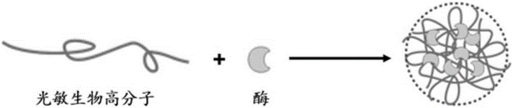
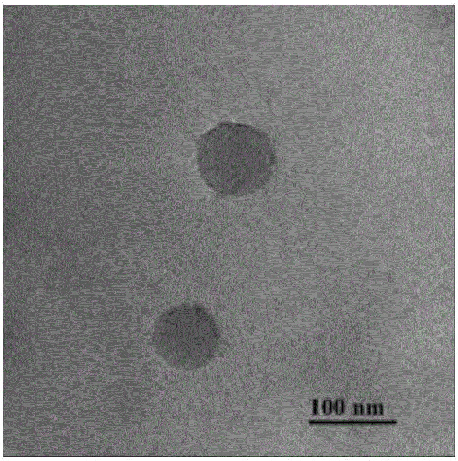
[0043] (2) Preparation of enzyme-loaded self-assembled nanoparticles
[0044] At room temperature, add 0.1 mg / mL glucose oxidase (GOx) solution dropwise into an equal volume of 0.5 mg / mL photosensitive biomacromolecul...
Embodiment 2
[0048] A method for preparing a biosensor based on photosensitive biopolymer-loaded enzymes, comprising the following specific steps:
[0049] (1) Preparation of photosensitive biopolymer
[0050] Biomacromolecule dextran (Dex), initiator 1-(3-dimethylaminopropyl)-3-ethylcarbodiimide hydrochloride and 4-dimethylaminopyridine, small molecule photosensitive monomer cinnamon The acid (CINN) was added dropwise into the dimethyl sulfoxide solvent, and reacted at 60°C for 5 hours to completely react the biomacromolecule with the photosensitive monomer; the resulting reactant solution was precipitated with precipitant ethanol and transferred to a dialysis bag for dialysis for 7 days To remove unreacted monomers and initiators; freeze-dry the purified polymer solution to obtain the photosensitive biomacromolecule Dex-CINN;
[0051] (2) Preparation of enzyme-loaded self-assembled nanoparticles
[0052] At room temperature, 1.0 mg / mL lactate oxidase (LOx) solution was added dropwise t...
Embodiment 3
[0056] A method for preparing a biosensor based on photosensitive biopolymer-loaded enzymes, comprising the following specific steps:
[0057] (1) Preparation of photosensitive biopolymer
[0058] Biomacromolecule polyglutamic acid (PGA), initiator 1-(3-dimethylaminopropyl)-3-ethylcarbodiimide hydrochloride and 4-dimethylaminopyridine, small molecule photosensitive monomer Hydroxyethyl methacrylate (HEMA) was added dropwise into the dimethyl sulfoxide solvent, and reacted at 25°C for 24 hours to completely react the biomacromolecule with the photosensitive monomer; Dialyze into a dialysis bag for 14 days to remove unreacted monomers and initiators; freeze-dry the purified polymer solution to obtain photosensitive biomacromolecule PGA-HEMA;
[0059] (2) Preparation of enzyme-loaded self-assembled nanoparticles
[0060] At room temperature, 10 mg / mL horseradish peroxidase (HRP) solution was added dropwise to 10 times its volume of 100 mg / mL photosensitive biomacromolecule PGA-...
PUM
| Property | Measurement | Unit |
|---|---|---|
| Particle size | aaaaa | aaaaa |
Abstract
Description
Claims
Application Information