

Microfluidic chip used for cell co-culture and cell culture method thereof
A microfluidic chip and cell culture technology, which is applied in the fields of biomedical engineering and cell biology research, can solve the problems of high reagent consumption, difficulty in high-throughput research, and inaccurate research results, and achieve simple processing and low cost. Shear force, the effect of simplifying the experimental procedure
- Summary
- Abstract
- Description
- Claims
- Application Information
AI Technical Summary
Problems solved by technology
Method used
Image

Examples
Embodiment 1
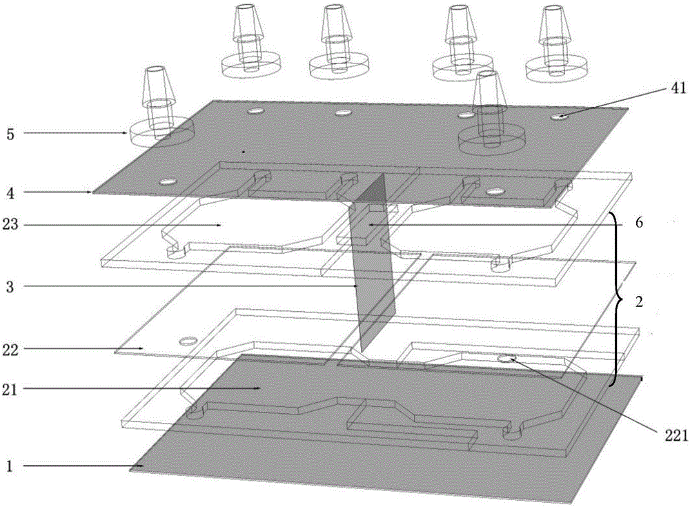
[0042] The invention provides a microfluidic chip for co-cultivation of cells, such as figure 1 As shown, the microfluidic chip includes a base layer 1, a cell culture chamber layer, 2, and an upper cover layer 4 from bottom to top. The cell culture chamber layer is composed of several cell culture chambers connected to each other on the same plane. For cell culture, Example 1 is an illustration of two cell culture chambers. The number of cell culture chambers in the present invention is not limited to 2, and can be formed by connecting multiple cell culture chambers.
[0043] The base layer can be a transparent material that has been treated with tissue culture according to the needs of the cultured cell type; or a transparent material that meets the requirements of cell culture through coating: such as polyethylene terephthalate PET, glass or polystyrene PS Wait.
[0044] The cell culture chamber layer is provided with a sieve 22 to divide each of the cell culture chambers ...
Embodiment 2
[0050] Preferably, an observation area is provided at the adjacent positions of the upper cover plate layer and the cell culture chamber layer corresponding to the two cell culture chambers, such as figure 1 As shown, the location of the observation area is not limited in the present invention, and all areas that can realize cell observation in two adjacent cell culture chambers are regarded as observation areas. The observation area can use any light-transmitting non-biologically toxic material as the upper cover to observe and detect the cells in two adjacent cell culture chambers.
Embodiment 3
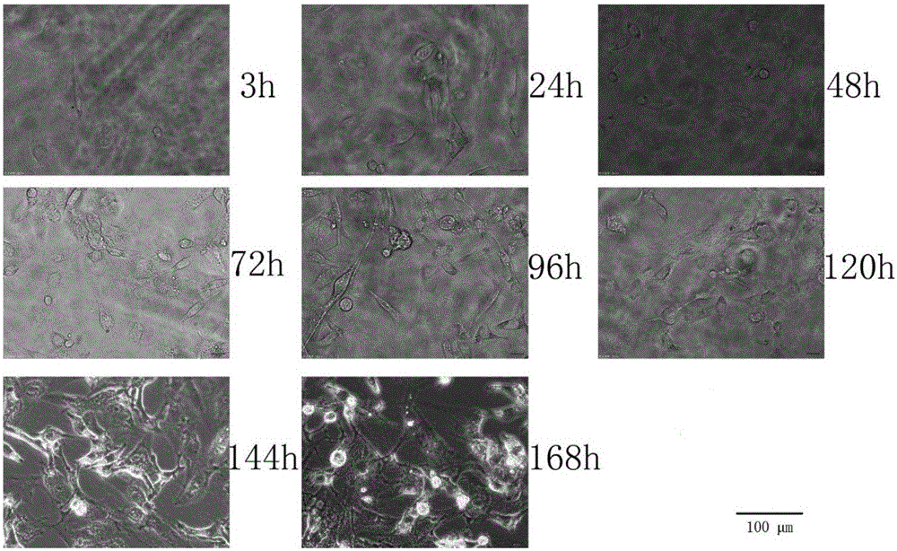
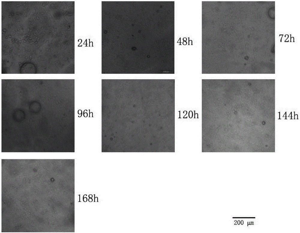
[0052] A microfluidic chip culture method for intermittent culture of cells co-cultivating human neuroblastoma SH-SY5Y and human glioma U87MG. In this embodiment, the structure of the microfluidic chip may not be provided with a filter Mesh and sieve to make microscopic imaging clear.
[0053] The method includes the following steps:
[0054] Human neuroblastoma SH-SY5Y and human glioma U87MG were frozen and stored at -150°C. Before the experiment, the cells were revived and placed at 37°C, 5% CO 2 In an incubator, culture in DMEM medium, supplemented with 10% fetal bovine serum, 100 U / mL penicillin G, 100 μg / mL streptomycin and 2% MEM NEAA (100X). The cells to be cultured by conventional cells grow to the logarithmic phase and then inoculated into the microfluidic chip.
[0055] The cells were digested with trypsin, and the digestion was terminated with fetal bovine serum after digestion. The digested cells were centrifuged at 1000rpm for 5min, and the culture medium was u...
PUM
| Property | Measurement | Unit |
|---|---|---|
| Aperture | aaaaa | aaaaa |
| Depth | aaaaa | aaaaa |
| Size | aaaaa | aaaaa |
Abstract
Description
Claims
Application Information