Chinese lung adenocarcinoma cell system and application thereof
A technique for lung adenocarcinoma cells and lung adenocarcinomas, applied in the field of microbial medicine, can solve problems such as ineffectiveness, and achieve the effect of promoting proliferation
- Summary
- Abstract
- Description
- Claims
- Application Information
AI Technical Summary
Problems solved by technology
Method used
Image
Examples
Embodiment 1
[0022] Isolation and processing of primary tumor cells
[0023] Fresh Chinese tumor tissue blocks obtained from the operating table of the hospital were isolated and cultured for primary tumor cells in a sterile environment.
[0024] 1. First put the tumor tissue block into a 50ml centrifuge tube, shake and wash 5 times with PBS containing 10 times the double antibody until there is no blood. Place the tissue block in a Petri dish and remove excess tissue, such as fat or dead tissue.
[0025] 2. Move the tissue to another plate, and use a cross scalpel to cut it into about 1mm 3 The size of the organization block. Take a 70um filter and place it on a 50ml centrifuge tube. Gently pressurize the core of the syringe to force the tissue through the mesh into the culture medium, draw the culture medium and blow it through the mesh to flush the cells down.
[0026] 3. Add 5ml of trypsin / collagenase II to the culture dish of the remaining tissue pieces, digest in a 37°C incubator...
Embodiment 2
[0032] Example 2. Monoclonal culture of primary tumor cells
[0033] 1. Digest the cells with trypsin to prepare a single cell suspension, count the cells, and dilute the cells to 1×10 cells / mL. The specific steps are as follows:
[0034] a) Dilute the digested cells to 1×10 5 individual / mL.
[0035] b) Take 1×10 5 cells / mL cell suspension 200uL, add to 20mL, become 1×10 3 individual / mL.
[0036] c) Divide 1×10 3 Cells / mL cell suspension 200uL, add to 20mL, become 1×10 cells / mL.
[0037] 2. Add 100uL of 1×10 cells / mL cell suspension to the microtiter culture plate of 96-well plate, and one colony will be formed in each well.
[0038] 3. A few hours after inoculation, find the culture well with only one cell through the microscope. The wells were marked and followed, and a drop in pH indicated cell growth, confirmed by microscopic observation. After the cells were overgrown, trypsinization was inserted into a 6-well plate for subculture.
Embodiment 3
[0039] Example 3. Detection and identification of Chinese lung adenocarcinoma cell line CAFQ1
[0040] 1. Cytomorphological observation of primary tumor cell lines
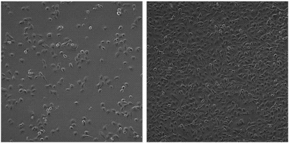
[0041] Under the light microscope, most of the tumor cells were spindle-shaped and oval-shaped, and a few were polygonal. The two poles of the cells were pointed, and the nucleus was obvious. The cells in the logarithmic growth phase spread well, and CAFQ1 randomly spread, and most of them were oval epithelioid cells, polygonal, and more regular in shape. Such as figure 1 shown.
[0042] 2. Immunofluorescence detection of cell marker proteins
[0043] 1) After trypsinizing the cells, suspend them in 1mL of complete culture medium, pipette fully to form a single-cell suspension, and perform cell counting. Insert 5×10 into 24-well plate as needed 4 cells. Before adding the cell suspension, add a small amount of medium to each well to make the slide and the culture dish stick together to reduce the entry of cel...
PUM
| Property | Measurement | Unit |
|---|---|---|
| Long trail | aaaaa | aaaaa |
Abstract
Description
Claims
Application Information
 - R&D
- Intellectual Property
- Life Sciences
- Materials
- Tech Scout
- Unparalleled Data Quality
- Higher Quality Content
- 60% Fewer Hallucinations
Browse by: Latest US Patents, China's latest patents, Technical Efficacy Thesaurus, Application Domain, Technology Topic, Popular Technical Reports.
© 2025 PatSnap. All rights reserved.Legal|Privacy policy|Modern Slavery Act Transparency Statement|Sitemap|About US| Contact US: help@patsnap.com


