Fluorescence bio-sensing method for detecting glutathione
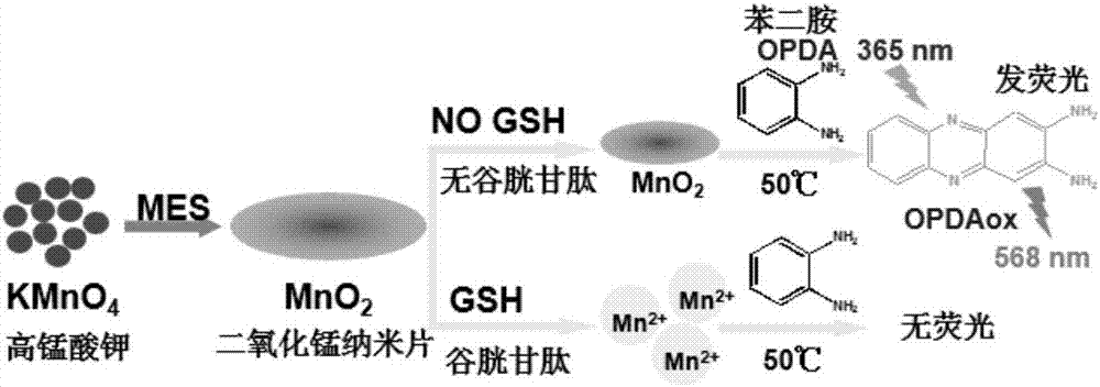
A glutathione and biosensing technology, applied in the field of fluorescent biosensing, can solve the problems of high cost, limited application, and many organic probe synthesis and purification steps, and achieves low cost, simple operation and high sensitivity. Effect
- Summary
- Abstract
- Description
- Claims
- Application Information
AI Technical Summary
Problems solved by technology
Method used
Image
Examples
Embodiment 1
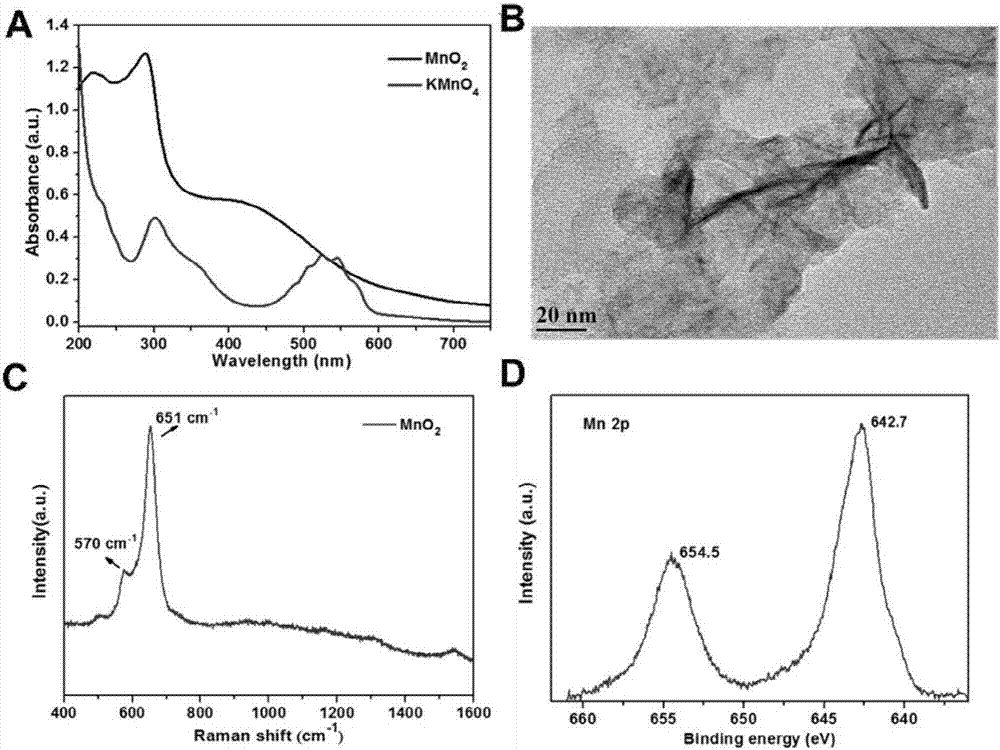
[0025] Synthesis of manganese dioxide nanomaterials: After mixing 1mL potassium permanganate solution (10mM) with 2.5mL MES buffer solution (0.1M, pH6.0), add 6.5mL secondary water; after that, place in an ultrasonic cleaner Medium ultrasonic reaction for 30 minutes until the formation of brown flocculent products. The brown product after the reaction was centrifuged at 8000 rpm for 10 minutes and washed 3 times with water to remove unreacted ions. Finally, the obtained manganese dioxide nanosheets were ultrasonically dispersed in secondary water at a concentration of 1 mg / mL. figure 2 It proves that the manganese dioxide material has been successfully synthesized. Among them, (A) is the absorption comparison chart of potassium permanganate solution and manganese dioxide after it is produced; (B) is the transmission electron microscope picture after synthesis, which proves that nanometer-sized Manganese dioxide nanosheets; (C) is the Raman spectrum of manganese dioxide nanos...
Embodiment 2
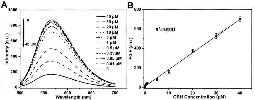
[0027] Fluorescent detection of glutathione: 2.5 μL of manganese dioxide nanosheets (1 mg / mL) and 200 μL of different concentrations of glutathione solutions (0, 10, 20, 30, 40 μM) were reacted at room temperature for 5 minutes; after that, 5 μL of 6 .8mM o-phenylenediamine (OPDA) solution was mixed and reacted in an oven or water bath at 50°C for 10 minutes. After cooling to room temperature, the fluorescence emission spectrum at 568nm was measured under the excitation of 420nm wavelength. When doing selectivity experiments, react different interfering substances with 2.5 μL of manganese dioxide nanosheets (1 mg / mL) at room temperature for 5 minutes; after that, add 5 μL of 6.8 mM o-phenylenediamine (OPDA) solution and mix well at 50 ° C. After reacting in oven or water bath for 10 minutes. After cooling to room temperature, test the fluorescence spectrum. Fluorescence response of glutathione as image 3 shown. The selectivity of the method for glutathione detection, as ...
Embodiment 3
[0029] Detection of glutathione in cell lysis solution: HeLa cells were cultured in RPMI 1640 medium containing 10% fetal bovine serum, penicillin (100 U / mL) and streptomycin (100 μg / mL) at 37°C in high humidity containing 5% in a carbon dioxide incubator. The grown HeLa cells were centrifuged, washed 3 times with cold PBS, and the cell concentration was calculated using a cell counter. Disperse the cell suspension into cold PBS so that each 100 μL solution contains 500, 5000, 10000, 20000, 30000, 40000 and other different numbers of cells, and place the cell suspension in an ice bath at 4 °C for 5 minutes sonication , after centrifugation at 10,000 rpm for 5 minutes, the supernatant was taken for testing. The supernatant was diluted to 200 μL and mixed with 2.5 μL of manganese dioxide nanosheets (1 mg / mL) for 5 minutes, then added 5 μL of 6.8 mM o-phenylenediamine (OPDA) solution and mixed, and placed in an oven or water bath at 50 ° C After 10 minutes of reaction. After c...
PUM
| Property | Measurement | Unit |
|---|---|---|
| concentration | aaaaa | aaaaa |
Abstract
Description
Claims
Application Information
 - R&D
- Intellectual Property
- Life Sciences
- Materials
- Tech Scout
- Unparalleled Data Quality
- Higher Quality Content
- 60% Fewer Hallucinations
Browse by: Latest US Patents, China's latest patents, Technical Efficacy Thesaurus, Application Domain, Technology Topic, Popular Technical Reports.
© 2025 PatSnap. All rights reserved.Legal|Privacy policy|Modern Slavery Act Transparency Statement|Sitemap|About US| Contact US: help@patsnap.com


