Fluorescent probe for bicolor differentiated imaging of dead and live cells and application of fluorescent probe
A fluorescent probe, life-and-death technology, applied in the direction of fluorescence/phosphorescence, analytical materials, luminescent materials, etc., can solve the problem of lack of fluorescent probes for two-color imaging of dead and alive cells, and achieve the effect of simple synthesis method and high yield
- Summary
- Abstract
- Description
- Claims
- Application Information
AI Technical Summary
Problems solved by technology
Method used
Image
Examples
Embodiment 1
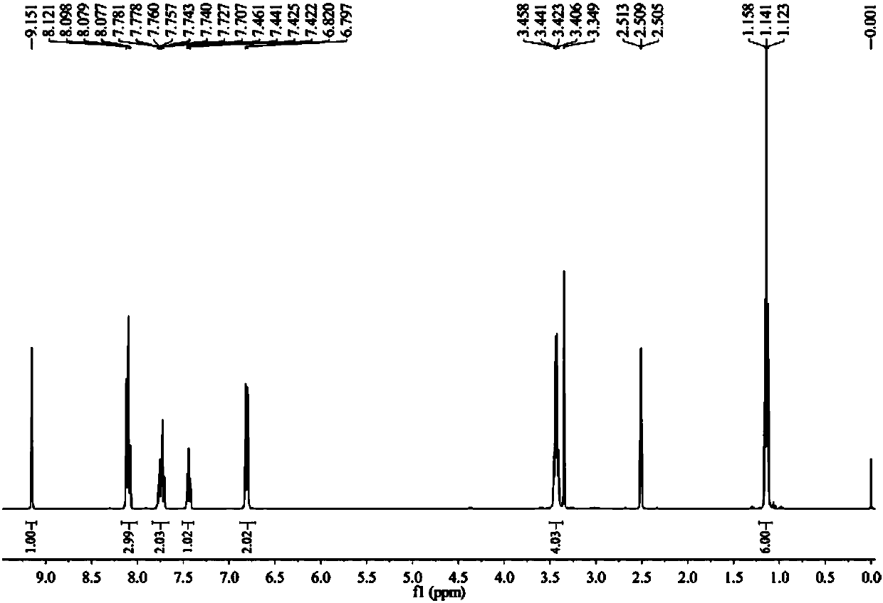
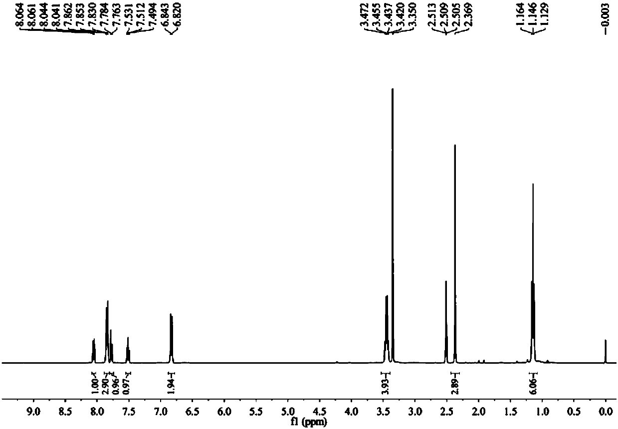
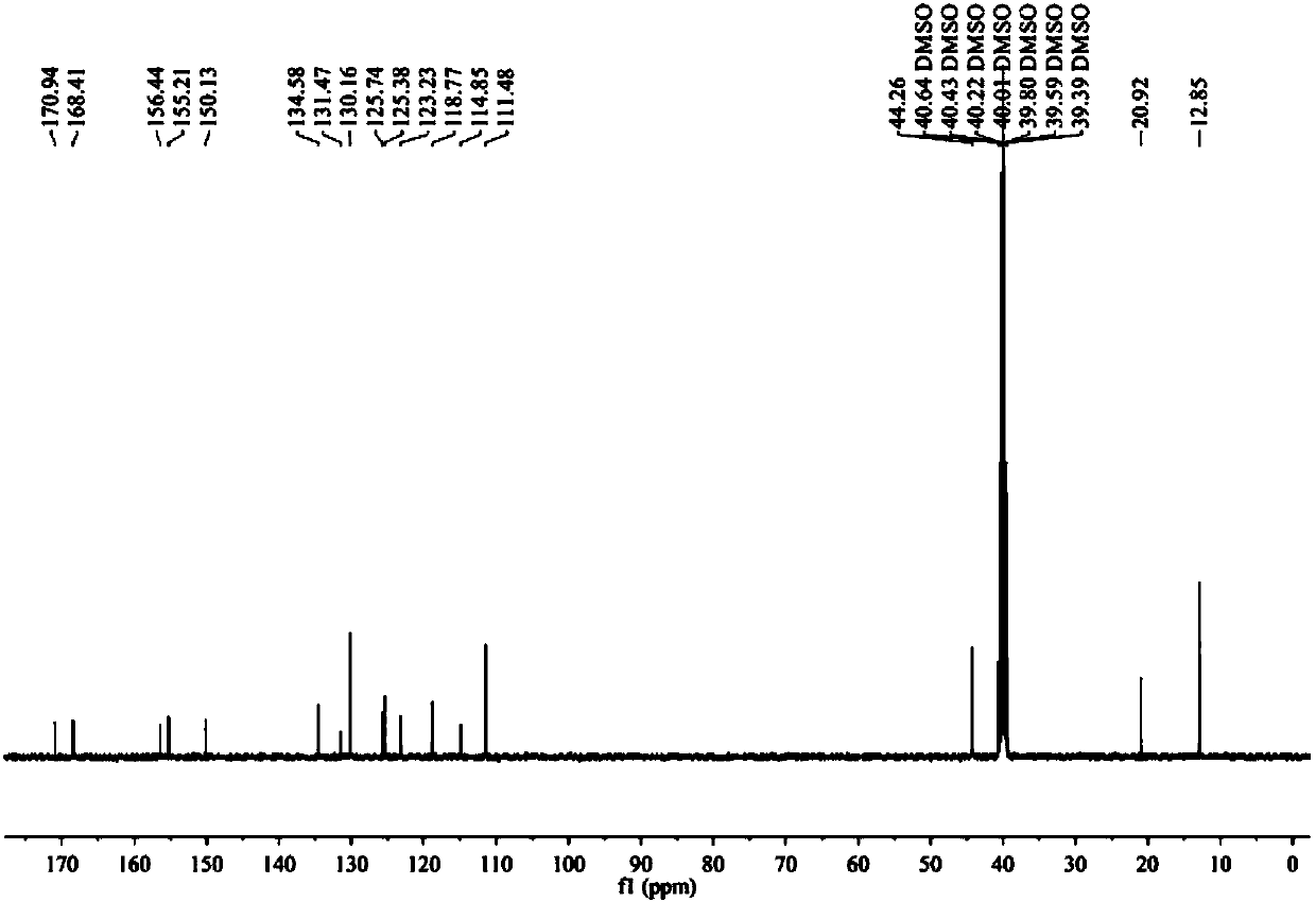
[0035] Example 1 Synthesis of fluorescent probes.
[0036] (1) Synthesis of 2-(4-diethylaminophenyl)-3-hydroxy-chromen-4-one (Compound 1).
[0037] Add 4-N,N-diethylaminobenzaldehyde (1.77 g, 10 mmol), o-hydroxyacetophenone (1.2 mL, 10 mmol) and 10 mL of ethanol into a round bottom flask. Sodium hydroxide solution (1.2 g, 30 mmol, 1 mL of water) was added with stirring. The reaction was stirred at room temperature for one day. After the reaction was completed, sodium hydroxide solution (0.4 g, 10 mmol, 1 mL of water) and 10 mL of hydrogen peroxide solution (30%) were added under ice-water bath conditions, stirred evenly, heated to 50° C. for 1 hour to complete the reaction, and cooled to room temperature. Pour the reaction liquid into 400mL water, neutralize it with sulfuric acid to neutral, a large amount of solids precipitated, filtered to obtain the crude product, and recrystallized in ethanol to obtain 1.51g of the pure product as orange crystals, with a yield of 50%. i...
Embodiment 2
[0040] The absorption spectrum test of embodiment 2 fluorescent probe DACA
[0041] The fluorescent probe DACA and the hydrolyzate compound 1 in Example 1 were weighed and prepared into a 5 mM mother solution with dimethyl sulfoxide (DMSO).
[0042] Take 10 μL of the probe mother liquor and the hydrolyzate mother liquor and add them to two identical 5mL volumetric flasks, add 1,4-dioxane to make up the volume, shake well and perform the absorption spectrum test, with the wavelength as the abscissa, and the absorbance and fluorescence Intensity is plotted on the ordinate as Figure 4 . Depend on Figure 4It can be seen that both the probe DACA and the hydrolyzate 1 have absorption at 405nm; under the excitation of 405nm light, the fluorescence peak of DACA is at 440nm, and the fluorescence peak of hydrolyzate 1 is at 570nm.
Embodiment 3
[0043] Example 3 Cell Imaging Research of Fluorescent Probe DACA
[0044] (1) True color fluorescence imaging
[0045] Set the density to 3×10 5 HeLa cells / mL were inoculated into sterilized 35mm imaging culture dishes in CO 2 Incubator (37°C, 5% CO 2 ) for more than 12 hours to allow the cells to adhere to the wall. The adherent cells were treated with 4% paraformaldehyde for 30 min to obtain dead cell samples. Dilute the mother solution described in Example 2 to 1 mM, and add the diluted solution to the dead and living cell culture dish so that the final concentration is 2 μM. Continue to culture under the same conditions for 40 minutes, then absorb the cell culture medium, wash the cells with phosphate buffered solution (PBS) for 3 times, use 405nm as the excitation wavelength, and use the spectral imaging module of the Nikon confocal microscope to collect the energy response Real-color imaging pictures of true fluorescent colors inside cells, such as Figure 5 shown....
PUM
 Abstract
Description
Claims
Application Information
 - R&D
- Intellectual Property
- Life Sciences
- Materials
- Tech Scout
- Unparalleled Data Quality
- Higher Quality Content
- 60% Fewer Hallucinations
Browse by: Latest US Patents, China's latest patents, Technical Efficacy Thesaurus, Application Domain, Technology Topic, Popular Technical Reports.
© 2025 PatSnap. All rights reserved.Legal|Privacy policy|Modern Slavery Act Transparency Statement|Sitemap|About US| Contact US: help@patsnap.com


