Bioglue for promoting blood vessel reconstruction after cell and organ transplantation and preparation method thereof

A technology of vascular reconstruction and biological glue, which is applied in prosthetics, medical science, etc., can solve the problems of reducing the potential risk of tumors induced by cell growth factors, prolonging the repair time, etc., achieving a simple and controllable preparation method and prolonging the tissue repair time , the effect of reducing potential risks

Active Publication Date: 2020-11-03
WENZHOU MEDICAL UNIV
View PDF7 Cites 0 Cited by
  • Summary
  • Abstract
  • Description
  • Claims
  • Application Information

AI Technical Summary

Problems solved by technology

At present, no effective technology has been seen, which can cooperate with cell growth factors to take effect in the transplantation of cells and organs. On the one hand, it prolongs the time for cell growth factors to play the role of promoting microvascular reconstruction and other tissue repair functions, and on the other hand, it reduces the potential of cell growth factors to induce tumors. risk

Method used


Image

  • Bioglue for promoting blood vessel reconstruction after cell and organ transplantation and preparation method thereof
  • Bioglue for promoting blood vessel reconstruction after cell and organ transplantation and preparation method thereof
  • Bioglue for promoting blood vessel reconstruction after cell and organ transplantation and preparation method thereof

Examples


Embodiment 1

[0025] The preparation of embodiment 1 bioglue

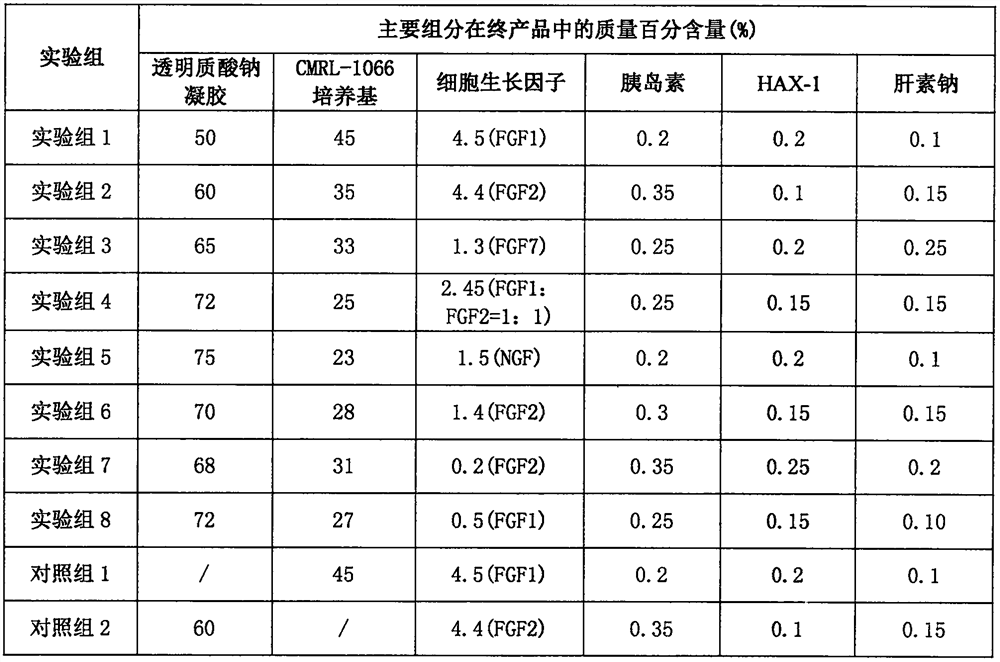
[0026] In this example, biological glues of each experimental group and control group were prepared according to the formula in Table 1, and the CMRL-1066 medium contained fetal bovine serum, penicillin, streptomycin and stabilizers. The preparation method of biological glue: add required cell growth factor, HAX-1, insulin and sodium heparin to sodium hyaluronate gel after sterilization (every 1000ml sodium hyaluronate gel contains 40g sodium hyaluronate and 10 g of phosphate buffered saline), mix well, add CMRL-1066 medium, mix well to form a semi-fluid gel, seal and place in a refrigerator at 4°C for 12 hours to remove air bubbles generated during the preparation process.

[0027] Table 1 Composition of each experimental group of bioglue

[0028]

[0029]

[0030] Note: " / " in the control group 1-7 means that the component is 0, and the component is replaced by water for injection, and the amount of water for injection ...

Embodiment 2

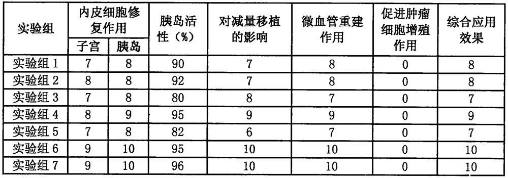
[0031] The application effect of embodiment 2 bioglue

[0032] In this example, the quality of each group of bioglue prepared in Example 1 was investigated through the following indicators.

[0033] (1) Repair function of endothelial cells

[0034]The biological glue prepared in each experimental group and control group in Example 1 was applied to the blood vessel suture after uterine transplantation in rats, and the tissue was taken two weeks later for HE staining and immunohistochemistry, etc., to investigate its effect on endothelial cell repair.

[0035] Mix the biological glue and mouse islets prepared by each experimental group and control group in Example 1 and co-culture them in vitro, collect cell samples every 4 hours, observe the internal structure of islet cells and the release of endocrine granules with a transmission electron microscope, and investigate its Effects on endothelial cell repair.

[0036] (2) Islet activity

[0037] Co-cultivate the biological glu...


PUM


Abstract

Biological glue promoting vascular reconstruction after cell and organ transplantation and a preparing method thereof are provided. Main components of the biological glue include sodium hyaluronate gel, a CMRL-1066 medium, a cell growth factor, HAX-1, insulin and heparin sodium. The biological glue effectively combines components synergistic with the cell growth factor to form a preparation, and the preparation is used for cell and organ transplantation to make an operating process more convenient, safer and more efficient, prolongs time for the cell growth factor to exert a function of promoting tissue repair, such as microvessel reconstruction, reduces the potential risk that the cell growth factor induces tumor. The biological glue is used for cell and organ transplantation and is particularly suitable for pancreas islet transplantation.

Description

technical field [0001] The invention relates to a bioglue and a preparation method thereof, in particular to a bioglue for promoting blood vessel reconstruction after cell and organ transplantation and a preparation method thereof. Background technique [0002] Sodium hyaluronate is a high-molecular polysaccharide substance containing uronic acid and hexosamine isolated from the vitreous body of the bull's eye. The molecular weight of sodium hyaluronate prepared by different processes is different. Sodium hyaluronate with natural properties The molecular weight is usually between 800,000 and 2.5 million. It is generally believed that the greater the molecular weight, the greater the elasticity and better adhesion. Sodium hyaluronate is a natural polysaccharide, and its gel has the characteristics of no species specificity, no toxicity, good biocompatibility and high solubility. As a new type of biological material, sodium hyaluronate gel is widely used in various clinical ...

Claims


Application Information

Patent Timeline
Patent Type & Authority Patents(China)
IPC IPC(8): A61L27/50A61L27/54A61L27/20A61L27/22
CPCA61L27/20A61L27/227A61L27/50A61L27/54A61L2300/236A61L2300/252A61L2300/412A61L2300/414C08L5/08C08L89/00
Inventor 赵应征傅红兴朱群燕刘成洋蒋煊鲁翠涛
Owner WENZHOU MEDICAL UNIV
Why Eureka
Try Eureka