3D-printing wound dressing material based on silk microsphere bio-ink and preparation method thereof

A technology of bio-ink and three-dimensional printing, which can be used in pharmaceutical formulations, microcapsules, drug delivery, etc., can solve the problems of low strength of fiber membranes, and achieve the effects of ensuring air permeability, wound coating, and uniformity

Active Publication Date: 2018-03-02
YANCHENG INST OF IND TECH
View PDF13 Cites 17 Cited by
  • Summary
  • Abstract
  • Description
  • Claims
  • Application Information

AI Technical Summary

Problems solved by technology

[0004] At present, most wound dressing materials are made of functional materials through electrospinning technology to form fiber membranes. The strength of the prepared fiber membranes is low, and most of them contain chemical cross-linking agents. Biocompatibility and long-term antibacterial properties need to be improved.

Method used


Image

  • 3D-printing wound dressing material based on silk microsphere bio-ink and preparation method thereof
  • 3D-printing wound dressing material based on silk microsphere bio-ink and preparation method thereof
  • 3D-printing wound dressing material based on silk microsphere bio-ink and preparation method thereof

Examples


Embodiment 1

[0033] (1) Centrifuge the aqueous solution of aspirin-coated silk protein microspheres with a particle size of 1-5 μm, remove the supernatant, add the aqueous solution of silk protein, stir ultrasonically, and mix uniformly to obtain a silk protein solution containing silk protein microspheres, wherein The mass fraction of the aspirin-coated fibroin microspheres is 3%, and the mass fraction of the silk protein is 5%.

[0034] (2) Add 1wt% ultraviolet photoinitiator Irgacure I-2959 and 3wt% polyvinyl alcohol ester to the silk protein solution containing silk protein microspheres, and mix evenly to obtain silk microsphere bio-ink.

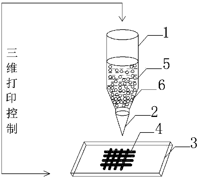
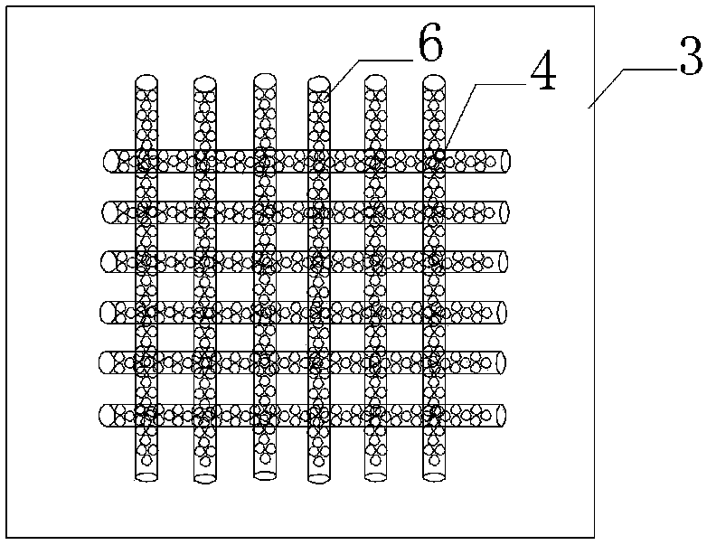
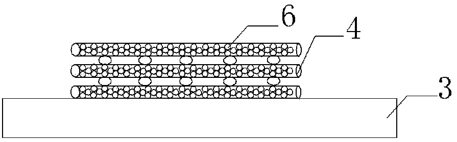
[0035] (3) Transfer the silk microsphere bio-ink to the syringe of the 3D printer, use cotton gauze as the base cloth for printing, establish a cyclical cross network structure scaffold model in MIMICS software, and adjust the needle cylinder pillow and cotton gauze The distance is 0.5mm, and the silk microsphere bio-ink is printed on the surface of ...

Embodiment 2

[0037] (1) Centrifuge the aqueous solution of aspirin-coated silk protein microspheres with a particle size of 1-5 μm, remove the supernatant, add the aqueous solution of silk protein, stir ultrasonically, and mix uniformly to obtain a silk protein solution containing silk protein microspheres, wherein The mass fraction of the aspirin-coated fibroin microspheres is 5%, and the mass fraction of the silk protein is 5%.

[0038] (2) Add 1wt% ultraviolet photoinitiator Irgacure I-2959 and 3wt% polyvinyl alcohol ester to the silk protein solution containing silk protein microspheres, and mix evenly to obtain silk microsphere bio-ink.

[0039] (3) The silk microsphere bio-ink was transferred to the syringe of the 3D printer, and cotton gauze was used as the base, and the bracket model of the cycled cross network structure was established in the MIMICS software, and the distance between the syringe pillow and the cotton gauze was adjusted as 0.5mm, the silk microsphere bio-ink was pr...

Embodiment 3

[0041] (1) Centrifuge the aqueous solution of aspirin-coated silk protein microspheres with a particle size of 1-5 μm, remove the supernatant, add the aqueous solution of silk protein, stir ultrasonically, and mix uniformly to obtain a silk protein solution containing silk protein microspheres, wherein The mass fraction of the aspirin-coated fibroin microspheres is 4%, and the mass fraction of the silk protein is 5%.

[0042] (2) Add 1wt% ultraviolet photoinitiator Irgacure I-2959 and 3wt% polyvinyl alcohol ester to the silk protein solution containing silk protein microspheres, and mix evenly to obtain silk microsphere bio-ink.

[0043] (3) The silk microsphere bio-ink was transferred to the syringe of the 3D printer, and cotton gauze was used as the base, and the bracket model of the cycled cross network structure was established in the MIMICS software, and the distance between the syringe pillow and the cotton gauze was adjusted as 0.5mm, the silk microsphere bio-ink is pri...


PUM

PropertyMeasurementUnit
particle diameteraaaaaaaaaa
wavelengthaaaaaaaaaa
thicknessaaaaaaaaaa

Abstract

The invention provides a 3D-printing wound dressing material based on silk microsphere bio-ink and a preparation method thereof. The preparation method includes the following steps: transferring silkmicrosphere bio-ink, which includes silk protein microspheres coating aspirin, silk protein water solution, ultraviolet initiator and polyvinyl alcohol ester, into a syringe of a 3D-printing machine;with a cotton gauze as a base printing cloth, printing the surface of the cotton gauze with the silk microsphere bio-ink according to a periodically circulated cross-network structure model; and rapidly curing and shaping the 3D-printing wound dressing material under ultraviolet irradiation. The product is based on 3D-printing technology, wherein the formed cross-network structure contains regularly arranged silk microspheres, and the silk microsphere bio-ink coating layer cures on the surface of the cotton gauze and does not completely penetrate through the cotton gauze. The 3D-printing wounddressing material has good mechanical performance and biocompatibility and excellent slow-release performance, can protect a wound for a long time and can accelerate healing of the wounding.

Description

technical field [0001] The invention belongs to the technical field of wound covering materials, in particular to a three-dimensional printing wound covering material based on silk microsphere bio-ink and a preparation method thereof. Background technique [0002] Three-dimensional printing technology, also known as three-dimensional rapid prototyping technology or additive weaving technology, is a new manufacturing technology. It is based on the principle of layered manufacturing and layer-by-layer superposition. The technology of rapidly weaving three-dimensional products of any complex shape through the accurate three-dimensional accumulation of materials based on computerized tomography and other data. [0003] At present, most of the inks used for 3D printing are biocompatible and biodegradable polymer materials. The commonly used ink materials are made of thermoplastics, silicone resin, collagen, gelatin or alginate, but these materials are affected by temperature and ...

Claims


Application Information

Patent Timeline
Patent Type & Authority Applications(China)
IPC IPC(8): A61L15/32A61L15/44A61L15/26A61L15/46
CPCA61L15/26A61L15/32A61L15/44A61L15/46A61L2300/21A61L2300/404A61L2300/412A61L2300/622C08L29/04C08L89/00
Inventor 王曙东马倩王可
Owner YANCHENG INST OF IND TECH
Why Eureka
Try Eureka