Novel staphylophage and composition, as well as preparation methods and application thereof

A technology of staphylococcus and bacteriophage, applied in the field of microorganisms

Active Publication Date: 2018-03-09
PHAGELUX (NANJING) BIO-TECH CO LTD
View PDF3 Cites 17 Cited by
  • Summary
  • Abstract
  • Description
  • Claims
  • Application Information

AI Technical Summary

Problems solved by technology

However, there are no relevant reports on virulent phages that can lyse Staphylococcus aureus and other staphylococci at the same time.

Method used


Image

  • Novel staphylophage and composition, as well as preparation methods and application thereof
  • Novel staphylophage and composition, as well as preparation methods and application thereof
  • Novel staphylophage and composition, as well as preparation methods and application thereof

Examples


Embodiment 1

[0067] Example 1: Isolation, preparation and purification of phage

[0068] In the present invention, the source samples of bacteriophage J1P1, J1P2, J1P3, J2-1P1 and J2-1P2 are collected in the cow dung piled up for 1 day and night in Jiangnan Dairy Farm, Jiangning District, Nanjing City, Jiangsu Province, and desorbed by 0.9% NaCl solution for 24 hours. Centrifuge at room temperature to get the supernatant, filter through double-layer filter paper, and then centrifuge at low speed at room temperature, then filter the supernatant with a 0.22 μm filter membrane.

[0069]Isolation of phage: Take 10ml of filtered supernatant, add it to 10ml of 2 times TSB medium, add 1ml of phage host bacteria logarithmic phase bacteria liquid at the same time, place it at 37°C for 16 hours, take the above culture, and put it under the condition of 8000rpm Centrifuge for 10 min, filter the supernatant with a 0.22 μm filter membrane, and set aside. Take 0.5ml of bacteriophage host bacteria logar...

Embodiment 2

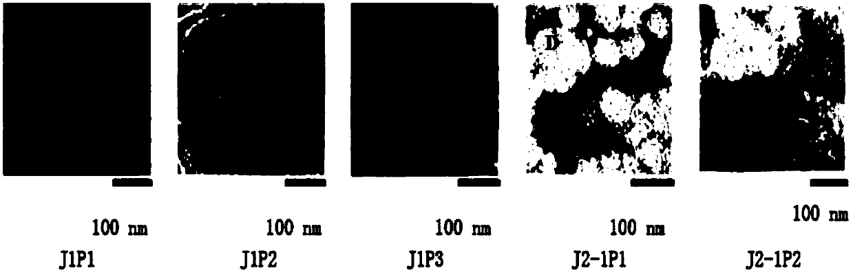
[0071] Embodiment 2: Electron microscope observation of phage

[0072] Take the supernatant of each phage culture obtained in Example 1 for electron microscope observation: take 20 μ l of the sample and drop it on the copper grid, wait for its natural precipitation for 15 minutes, absorb excess liquid from the side with filter paper, add 1 drop of 2% phosphotungstic acid (PTA ) on a copper grid, dyed for 10 minutes, sucked the dye solution from the side with filter paper, and observed with an electron microscope after drying: the results are as follows: figure 1 As shown, it can be seen through the transmission electron microscope that the morphology of the J1P1 phage is observed under the electron microscope, and it has a regular polyhedral head structure and a scalable tail. The diameter of the head is about 100nm, the length of the tail is about 200nm, and the end of the tail has six Short spines; J1P2 phage morphology was found to have a regular polyhedron head structure a...

Embodiment 3

[0073] Example 3: Extraction and sequencing of phage genome

[0074] Take 100ml of each phage prepared in Example 1, add DNaseI and RNaseA at a final concentration of 1 μg / ml, incubate at 37°C for 60 minutes, add 5.84g Nacl (final concentration 1mol / L), dissolve and place in an ice bath for 1 hour. Centrifuge at 11,000 rpm for 10 min at 4°C, and transfer the supernatant to a new centrifuge tube. Add solid PEG8000 (final concentration 10%, that is, add 10g to 100ml), and after it is completely dissolved, put it in ice bath for at least 1h. Centrifuge at 11,000 rpm for 20 min at 4°C, and resuspend the pellet with a small amount of SM solution. Add an equal volume of chloroform and isoamyl alcohol for extraction, shake gently for 30 s, centrifuge at 8000 rpm for 1 min, absorb the supernatant, and repeat the extraction until clarification. Add DNase I and RNase A again to a final concentration of 1 μg / ml, and react at 37°C for 30-60 minutes. Add EDTA to a final concentration of...


PUM

PropertyMeasurementUnit
molecular weightaaaaaaaaaa
diameteraaaaaaaaaa

Abstract

The invention relates to the field of microorganisms, and specifically provides a novel staphylophage and a composition, as well as preparation methods and application thereof. The phage is aureus staphylophage J1P1 with the storage number of CCTCC NO: M2016284, aureus staphylophage J1P2 with the storage of CCTCC NO: M2016285, aureus staphylophage J1P3 with the storage of CCTCC NO: M2016286, aureus staphylophage J2-1P1 with the storage of CCTCC NO: M2016287, or aureus staphylophage J2-1P2 with the storage of CCTCC NO: M2016288. The phage is capable of preventing and treating human or animal bacterial infectious diseases caused by various staphylococcocci, particularly by aureus staphylophage; a good phage resource is provided to develop novel antimicrobial preparations; the novel staphylophage has a good application and development prospect.

Description

technical field [0001] The invention belongs to the technical field of microorganisms, and in particular relates to novel staphylococcal bacteriophages, their composition, their preparation method and application. Background technique [0002] The genus Staphylococcus belongs to the class Bacilli, the order Bacillales, and the family Staphylococcaceae. Staphylococcus is widely distributed in the air, feed, drinking water, ground and surface of objects. There are also parasites in the skin, mucous membranes, intestinal tract, respiratory tract and mammary glands of humans and livestock, and they are the main pathogenic bacteria that cause many bacterial diseases in humans and livestock. In 2004, "Burger Handbook of Systematic Bacteriology" divided this genus into 37 species, many of which were also divided into subspecies. According to its ability to coagulate rabbit plasma, it can be divided into two categories: coagulase-positive bacteria and coagulase-negative bacteria. ...

Claims


Application Information

Patent Timeline
Patent Type & Authority Applications(China)
IPC IPC(8): C12N7/00A61K35/76A61P31/04A61P17/00A61P15/00
CPCA61K35/76C12N7/00C12N2795/00021
Inventor 肖逍苏胜兵费文斌凌文乔欢丛郁徐旭凌周思翔熊剑胜霍茨蒙德·曼德维尔沈婵娟
Owner PHAGELUX (NANJING) BIO-TECH CO LTD
Why Eureka
Try Eureka