Preparation method of a transplantable scaffold for inducing circulating tumor cell metastasis

A technology of tumor cells and induced circulation, which is applied in the direction of drug devices and other medical devices, can solve the problems of exosomes that cannot be used in structure, drug loading and sustained release, and bioactive molecule inactivation, etc., to achieve non-degradability Good, overcoming the difficulty of transplantation, good denaturation and mechanical properties

Active Publication Date: 2019-08-20
杭州多泰科技有限公司
View PDF4 Cites 0 Cited by
  • Summary
  • Abstract
  • Description
  • Claims
  • Application Information

AI Technical Summary

Problems solved by technology

However, the current scaffold materials that meet such requirements, such as silica gel materials, are relatively dense in structure and cannot be used for the loading and sustained release of exosomes and drugs, and the preparation process can easily inactivate biologically active molecules.

Method used


Image

  • Preparation method of a transplantable scaffold for inducing circulating tumor cell metastasis

Examples


Embodiment 1

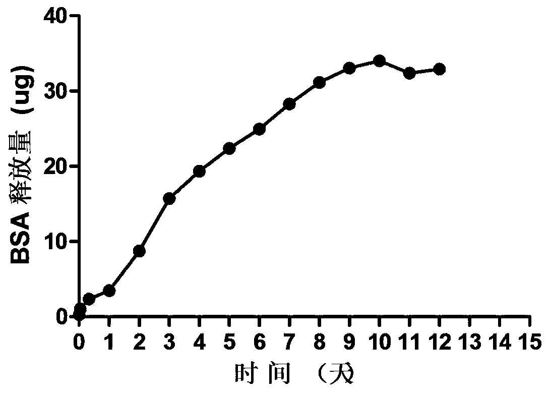
[0030] see figure 1 , mix PDMS and curing agent according to 10:1, drop it on the cover glass, put it on the coating machine and spin coat it for 90s, the speed is 400rpm / min, heat at 60°C for 20min, and the PDMS is cured; the cured PDMS is sprayed with plasma The exosomes were treated for 1 min; the polyallylamine hydrochloride solution was added dropwise on the surface of the PDMS membrane, 400rpm / min spin-coated for 90s, then water was added dropwise, 2000rpm / min spin-coated for 60s, and then the PBS solution containing exosomes was sprayed, and then added dropwise Polystyrene sulfonate aqueous solution was spin-coated at 400rpm / min for 90s, then water was added dropwise, and spin-coated at 2000rpm / min for 60s, and this was cycled 20 times to form layer-by-layer self-assembled membranes loaded with exosomes on the PDMS surface. Wherein polystyrene sulfonate, polypropylene amine hydrochloride aqueous solution, concentration 5g / L, add 0.15M sodium chloride, adjust pH value to...

Embodiment 2

[0033] Mix PDMS and curing agent 15:1, drop it on the cover glass, put it on the coating machine and spin coat it for 30s, the rotation speed is 5000rpm / min, heat at 120°C for 20min, and the PDMS is cured; the cured PDMS is treated with plasma 10min; Add polylysine solution dropwise on the surface of PDMS membrane, spin-coat at 3000rpm / min for 40s, then add water dropwise, spin-coat at 3000rpm / min for 40s, then spray PBS solution containing exosomes, then add hyaluronic acid aqueous solution dropwise, Spin-coat at 3000rpm / min for 40s, then add water dropwise, spin-coat at 3000rpm / min for 40s, cycle 100 times, and form layer-by-layer self-assembled films loaded with exosomes on the surface of PDMS. Wherein hyaluronic acid, polylysine aqueous solution, concentration 1g / L, add 0.15M sodium chloride, adjust pH value to be 3.

[0034] The PDMS membrane was peeled off from the substrate, sterilized with hexane oxide gas, and implanted into the omentum fat pad.

Embodiment 3

[0036] Mix PDMS and curing agent 10:1, drop it on the cover glass, put it on the coating machine and spin coat it for 90s, the rotation speed is 3000rpm / min, heat at 100°C for 20min, and the PDMS is cured; the cured PDMS is treated with plasma 5min; Add chitosan aqueous solution dropwise on the surface of PDMS membrane, spin-coat at 4000rpm / min for 20s, then add water dropwise, spin-coat at 4000rpm / min for 20s, then spray PBS solution containing exosomes, then add hyaluronic acid aqueous solution dropwise, 4000rpm / min spin coating for 20s, then add water dropwise, 4000rpm / min spin coating for 20s, and cycle 50 times in this way to form layer-by-layer self-assembled films loaded with exosomes on the surface of PDMS. Wherein hyaluronic acid, chitosan aqueous solution, concentration 2g / L, add 0.15M sodium chloride, adjust pH value to be 3.

[0037] The PDMS membrane was peeled off from the substrate, sterilized with hexane oxide gas, and implanted into the omentum fat pad.


PUM


Abstract

The invention discloses a preparation method of an implantable stent capable of inducing metastasis of circulating tumor cells. Specifically, the preparation method comprises the following steps: spin-coating PDMS, so that a thin film is formed, and curing the thin film; conducting plasma treatment on the obtained PDMS film, so that the surface of the PDMS film is negatively charged; dropping a cationic polymer solution onto the surface of the PDMS film, implementing spin-coating, then dropping clean water and implementing spin-coating, then spraying a PBS solution containing an exosome, thendropping an anionic polymer solution, implementing spin-coating, then dropping clean water and implementing spin-coating, therefore through such circulation, a layer-by-layer self-assembled film containing the exosome is loaded on the PDMS surface; and removing the PDMS from a substrate, and implementing disinfecting via epoxy hexane gas. The implantable stent provided by the invention, as a flexible thin film, is excellent in denaturation capacity and mechanical performance, and implanting difficulty, caused by space limitation, can be overcome; and the PDMS is modified via a layer-by-layer self-assembling technology, so that biocompatibility, non-degradability and excellent deformation capacity and mechanical performance are reserved, and meanwhile, loading and sustained release of the exosome / drugs are achieved.

Description

technical field [0001] The invention relates to the technical field of tissue engineering, in particular to a method for preparing a transplantable scaffold for inducing the transfer of circulating tumor cells. Background technique [0002] Complications caused by metastasis of malignant tumors to functional organs (lung, liver, brain, etc.) are the main cause of tumor death. Circulating tumor cells (CTCs) are a type of tumor cells released from solid tumors or metastases into the peripheral blood circulation. Existing studies have proved that they play an important role in postoperative recurrence and distant metastasis of malignant tumors. However, the current mainstream tumor treatment methods, such as chemotherapy, radiotherapy, and surgical resection, are difficult to completely eliminate these circulating tumor cells. [0003] Exosomes are nanovesicles secreted by cells with a lipid bilayer structure, with a particle size distribution of 30-100 nm, and are widely dist...

Claims


Application Information

Patent Timeline
Patent Type & Authority Patents(China)
IPC IPC(8): A61M31/00
Inventor 王娇
Owner 杭州多泰科技有限公司
Why Eureka
Try Eureka