Microcystis toxin immune quantification test strip based on fluorescent microsphere mark
A technology of microcystin and fluorescent microspheres, which is applied in the direction of measuring devices, instruments, scientific instruments, etc., can solve the problems of wide range of microcystin pollution, detection methods that cannot meet the requirements, and narrow dynamic detection range. The effect of market demand, small error and high sensitivity
- Summary
- Abstract
- Description
- Claims
- Application Information
AI Technical Summary
Problems solved by technology
Method used
Image
Examples
Embodiment 1
[0035] Example 1 Fluorescent probe preparation of Eu-fluorescent microspheres labeled microcystin artificial antigen (or goat anti-rabbit secondary antibody)
[0036] The specific preparation method is as follows:
[0037] (1) Take 50 μL (1% solid content) of carboxyl fluorescent microspheres (purchased from Xiamen Nodao Technology Co., Ltd., particle size 300 nm) placed at 4°C, ultrasonically disperse, and add 600-800 μL of 2-(N-morphine at a concentration of 0.05M Phenyl)ethanesulfonic acid (MES, C 6 h 13 NO 4 S·H 2 O) Activation buffer, centrifuged at 16500rpm for 10-15min (the temperature is controlled at about 15°C during centrifugation);
[0038] (2) Discard the supernatant, add 600-800μL MES buffer, resuspend by ultrasonication, and repeat centrifugal washing 2-3 times;
[0039] (3) Discard the supernatant, add 200 μL MES buffer for ultrasonic resuspension, add 50 μL 10 mg / mL 1-(3-dimethylaminopropyl)-3-ethylcarbodiimide hydrochloride (EDC, C 8 h 17 N 3 HCl), 50...
Embodiment 2
[0045] Example 2 Assembly of Microcystin Immunity Test Strips Based on Labeled Fluorescent Microspheres
[0046] The structural diagram of the microcystin immune test strip is as follows: image 3 As shown, there are sample pads, nitrocellulose (NC) membrane and absorbent paper from left to right on the bottom plate. The key to test strip assembly is to ensure consistent transmissibility between each part. The sample pad is stacked on the NC. On the membrane, the two overlap by about 5mm. Similarly, the absorbent paper is stacked on the NC membrane, and the two overlap by about 5mm. Use a strip cutter to cut the pasted board into test strips about 4mm wide. Assemble and store sealed at 4°C for later use.
[0047] The assembly method is as follows: Spray goat anti-mouse secondary antibody (1mg / mL) on the nitrocellulose membrane as the detection line (T line), and spray rabbit IgG antibody (1mg / mL) on the nitrocellulose membrane as the quality control line (C line), the sprayi...
Embodiment 3
[0048] Example 3 Drawing of the Standard Curve Based on the Microcystin Immunization Test Strip of Labeled Fluorescent Microspheres
[0049] The method of drawing the standard curve is:
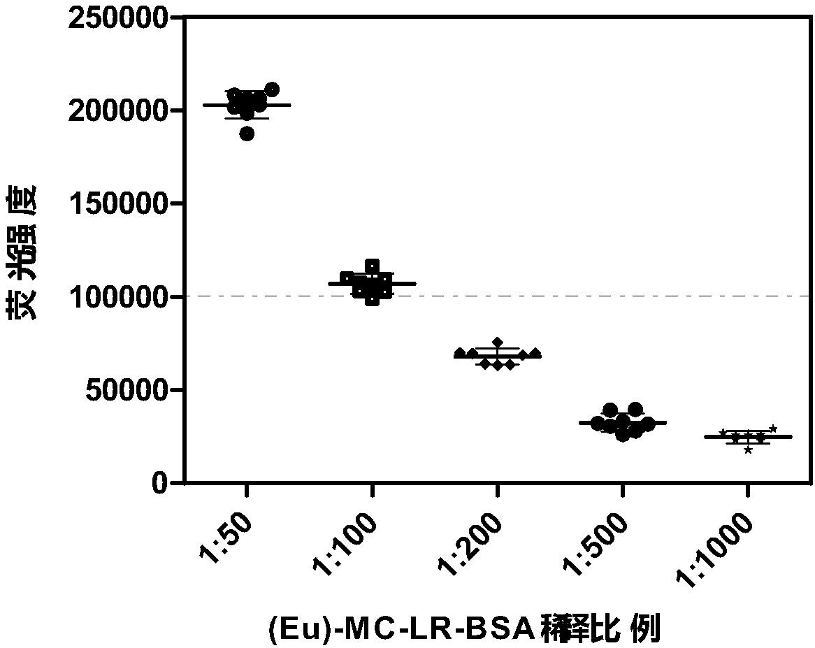
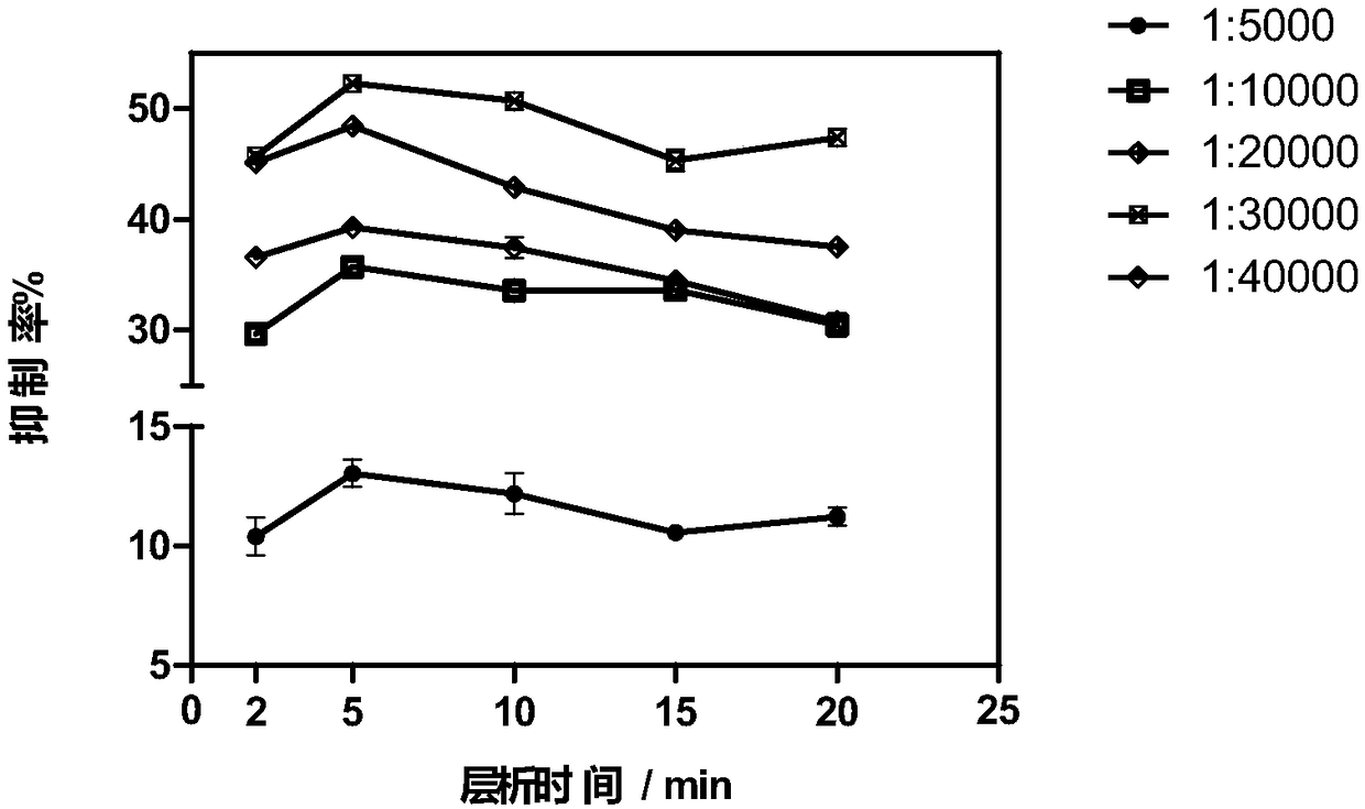
[0050] Eu-fluorescent microsphere-labeled microcystin artificial antigen (Eu-MC-LR-BSA) and goat anti-rabbit secondary antibody (Eu-Goat-anti-Rabbit) were used as fluorescent probes, and 30 μL of microcystin Standards, 30 μL of Eu-MC-LR-BSA, 30 μL of microcystin monoclonal antibody ascites and 5 μL of Eu-Goat-anti-Rabbit were added dropwise to a 96-well plate, shaken at room temperature for 10 minutes, and 70 μL of the mixture was slowly dropped into the test strip sample Pad, chromatographed at 37°C for 5min. Then record T value and C value by HG-98 immunological quantitative analyzer. The concentration gradient of microcystin standard substance is: 0μg / L, 0.1μg / L, 0.25μg / L, 0.5μg / L, 1μg / L, 2μg / L, 5μg / L. Take the logarithmic value of the standard substance concentration as the abscissa, a...
PUM
 Abstract
Description
Claims
Application Information
 - R&D
- Intellectual Property
- Life Sciences
- Materials
- Tech Scout
- Unparalleled Data Quality
- Higher Quality Content
- 60% Fewer Hallucinations
Browse by: Latest US Patents, China's latest patents, Technical Efficacy Thesaurus, Application Domain, Technology Topic, Popular Technical Reports.
© 2025 PatSnap. All rights reserved.Legal|Privacy policy|Modern Slavery Act Transparency Statement|Sitemap|About US| Contact US: help@patsnap.com


