

Joint detection kit for testing intestinal mucosa injury and preparation method of joint detection kit
A joint detection technology for intestinal mucosal injury, applied in the direction of measuring devices, instruments, scientific instruments, etc., can solve the problems of inability to perform single-person detection in time, narrow detection linear range, and inaccurate detection results, etc., to achieve long-lasting luminous signals, Effect of improving detection sensitivity and improving linear range
- Summary
- Abstract
- Description
- Claims
- Application Information
AI Technical Summary
Problems solved by technology
Method used
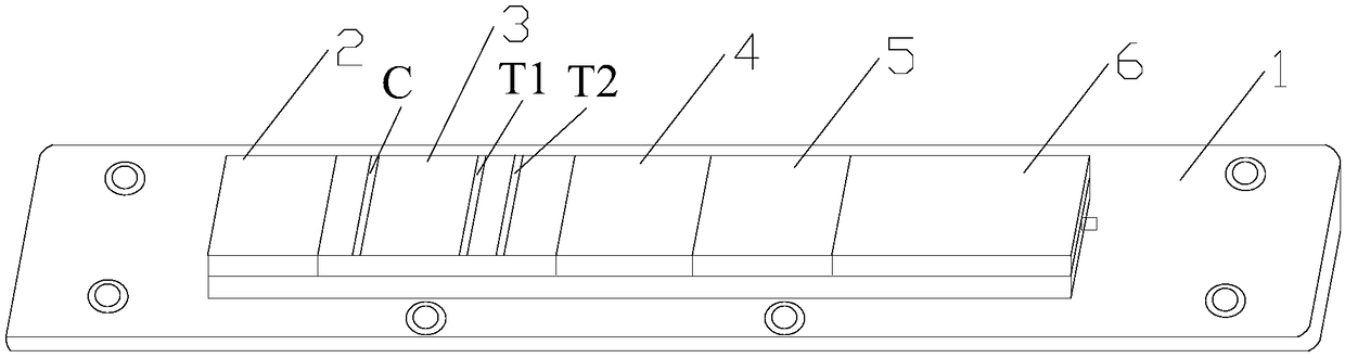
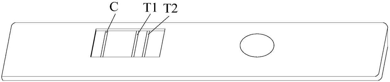
Image

Examples
Embodiment 1
[0034] 1. Prepare the mixture of quantum dot-labeled calprotectin antibody 1
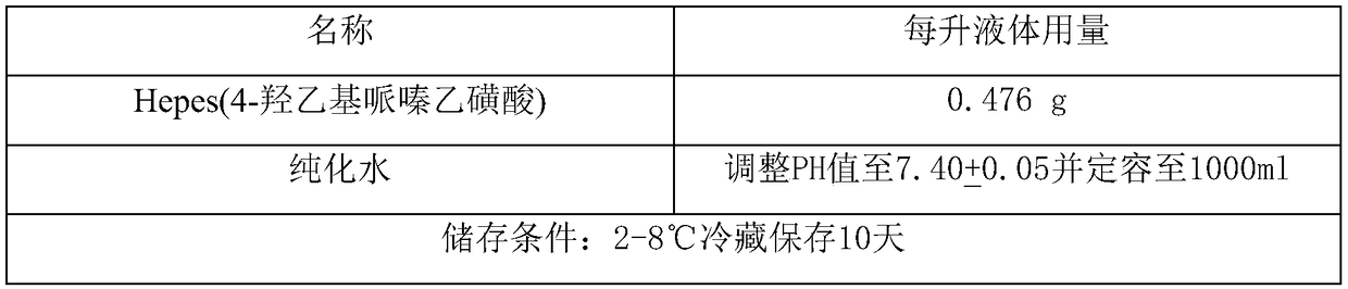
[0035] (1) Take 1ml of quantum dot fluorescent microspheres (the concentration is 10mg / ml, the particle size is 180nm, CdSe / ZnS, the surface group is carboxyl group, the excitation wavelength is 410, the emission wavelength is 615, purchased from: Suzhou Weidu Biotechnology Co., Ltd. ), adding 16.5ml of labeling buffer, and ultrasonically mixing;
[0036] labeling buffer
[0037]
[0038](2) Add 0.5ml each of Activation Buffer 1 and Activator Buffer 2, shake and react at 37°C for 25 minutes, and ultrasonically disperse for 20 seconds every 4 minutes;
[0039] Activation buffer 1
[0040]
[0041] Activation buffer 2
[0042]
[0043] (3) Add 1.4 mg of Calprotectin Antibody 1 (purchased from: Medix Biochemica), ultrasonically mix, shake and react at 37°C for 1 hour and 20 minutes, and ultrasonically disperse for 25 seconds every 8 minutes during this period;
[0044] (4) Add 1.5ml blocki...
Embodiment 2
[0095] 1. Prepare the mixture of quantum dot-labeled calprotectin antibody 1
[0096] (1) Take 1ml of quantum dot fluorescent microspheres (the concentration is 10mg / ml, the particle size is 160nm, InP, the surface group is carboxyl, the excitation wavelength is 405, the emission wavelength is 605), add 15.5ml of labeling buffer, and mix well by ultrasonic ;
[0097] labeling buffer
[0098]
[0099] The quantum dot fluorescent microspheres can also be silicon, germanium, cadmium sulfide, cadmium telluride, zinc selenide, lead sulfide, lead selenide, indium arsenide and polystyrene microspheres, polymethacrylate microspheres, poly Compounding of ethylene toluene microspheres or silica microspheres;
[0100](2) Add 0.45ml each of Activation Buffer 1 and Activation Buffer 2, shake and react at 37°C for 30 minutes, and ultrasonically disperse for 30 seconds every 5 minutes during this period;
[0101] Activation buffer 1
[0102]
[0103]
[0104] Activation buffer 2...
PUM
| Property | Measurement | Unit |
|---|---|---|
| Particle size | aaaaa | aaaaa |
| Particle size | aaaaa | aaaaa |
| Sensitivity | aaaaa | aaaaa |
Abstract
Description
Claims
Application Information