Two-photon red-light-emitting fluorescent probe for detecting mitochondria

A fluorescent probe and two-photon excitation technology, applied in fluorescence/phosphorescence, luminescent materials, measurement devices, etc., can solve problems that need to be developed, and achieve the effects of small light damage, deep penetration depth, and low photobleaching effect

Inactive Publication Date: 2018-11-16
UNIV OF JINAN
View PDF1 Cites 6 Cited by
  • Summary
  • Abstract
  • Description
  • Claims
  • Application Information

AI Technical Summary

Problems solved by technology

And so far, the report of this kind of molecule is still less, to be developed

Method used


Image

  • Two-photon red-light-emitting fluorescent probe for detecting mitochondria
  • Two-photon red-light-emitting fluorescent probe for detecting mitochondria
  • Two-photon red-light-emitting fluorescent probe for detecting mitochondria

Examples


Embodiment 1

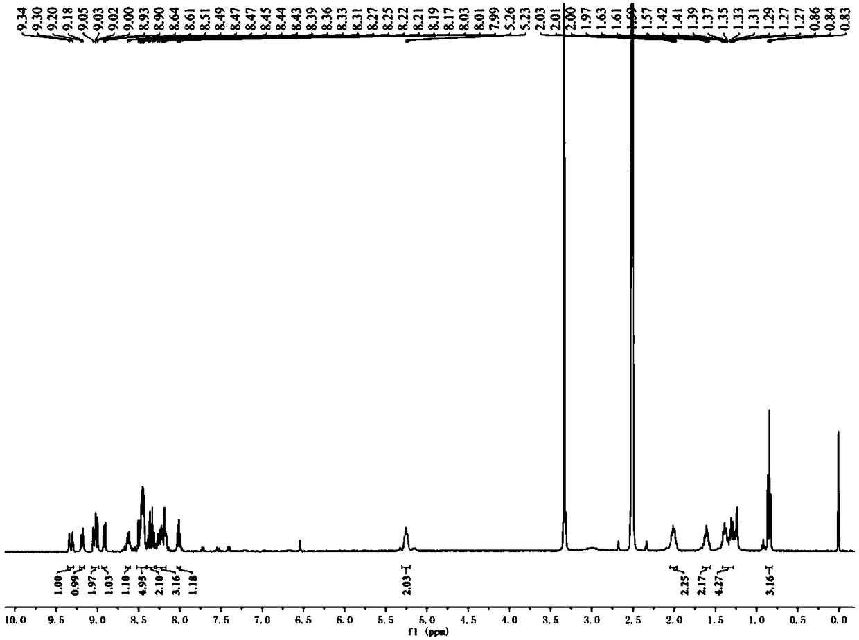
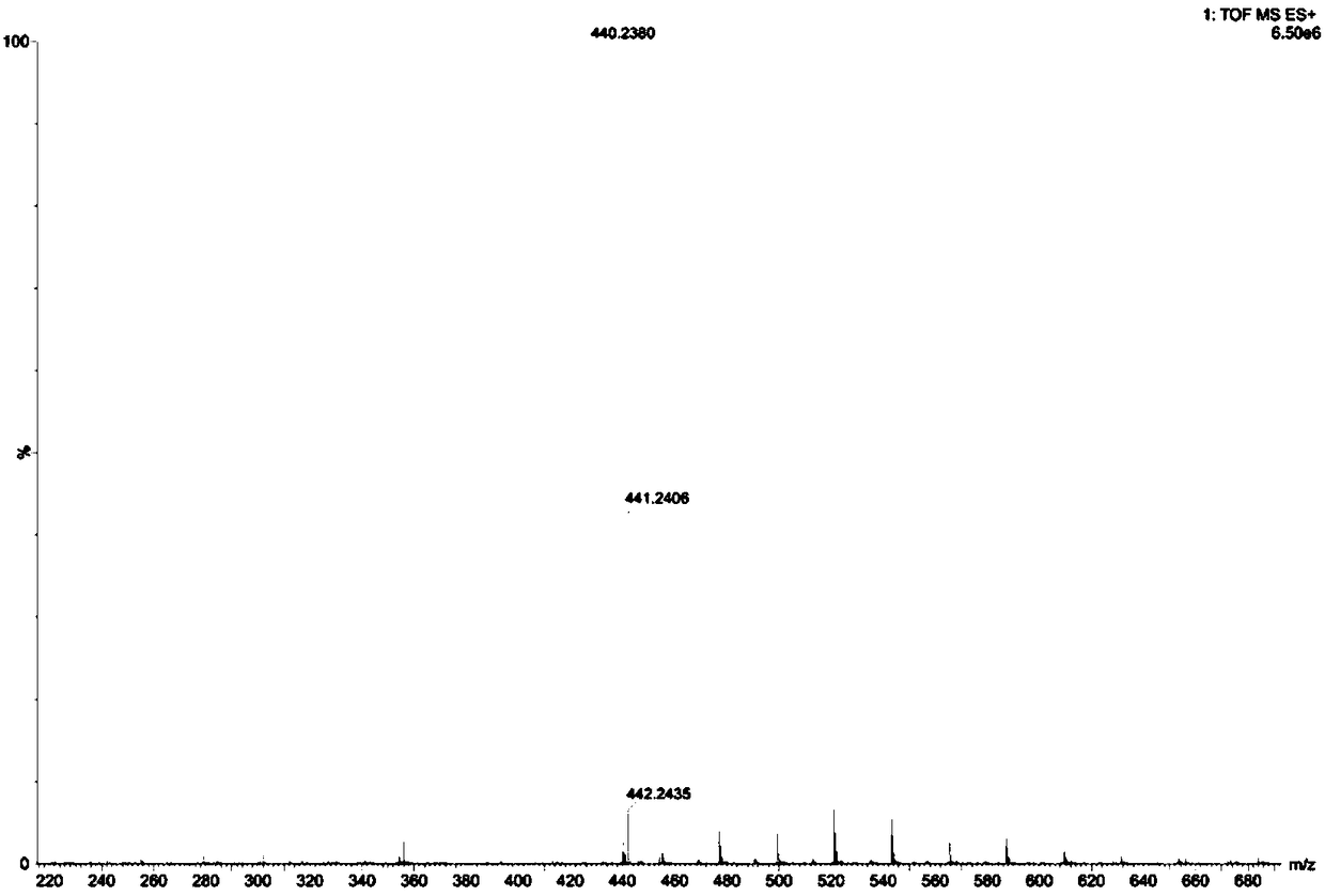
[0028] Example 1 Synthesis of Fluorescent Probe HVQ

[0029] Use ethanol as solvent, add 2-methylquinoline, add 1.2 eq iodohexane solution, heat to 80°C for 24 h, after the reaction is completed, cool the reaction system to room temperature, add 1 eq 1-pyrene carboxaldehyde, add 1 eq of pyrrolidine, stirred at room temperature for 12 hours, a large amount of solids precipitated, and the crude product was obtained by filtration. The crude product was redone with absolute ethanol three times, and then recrystallized in ethanol to obtain the pure product 2-(pyrene-1-vinyl )-N-hexyl-quinoline iodonium salt, the productive rate is 48%. product of 1 The H NMR and HRMS spectra are as follows figure 1 with figure 2 Shown: 1 H NMR (400 MHz, DMSO- d 6 ) δ 9.32 (d, J = 15.2 Hz, 1H), 9.19 (d, J = 8.9 Hz, 1H), 9.03 (dd, J = 12.8, 9.1 Hz, 2H), 8.92 (d, J = 8.3 Hz, 1H), 8.63 (d, J = 9.1 Hz, 1H), 8.52 – 8.42 (m, 5H), 8.39 – 8.30 (m, 2H), 8.28 -8.17 (m, 3H), 8.01 (t, J = 7.5 ...

Embodiment 2

[0030] Example 2 Cell Imaging of Fluorescent Probe HVQ

[0031] (1) Cell culture, treatment and staining

[0032] Set the density to 3×10 5 cells / mL of HeLa cells were inoculated into sterile 35 mm imaging culture dishes in CO 2 Incubator (at 37°C, 5% CO 2 ) for more than 12 hours to allow the cells to adhere to the wall. Adherent cells were treated with 4% paraformaldehyde for 30 min to obtain dead cell samples. The DMSO solution of the fluorescent probe prepared in Example 1 with a concentration of 2.5 mM was prepared as the mother solution, and the mother solution was added to the dead and living cell culture dish so that the final concentration was 5 μM. Continue to culture under the same conditions for 1 h, then aspirate the cell culture medium, wash the cells with the medium for 3 times, and then perform the cell imaging experiment.

[0033] (2) Confocal microscope imaging

[0034] The excitation wavelengths were 405 nm and 800 nm respectively, and the collection ...

Embodiment 3

[0035] Example 3 Co-localization of probes and commercial probes in living cells

[0036] In order to further confirm the staining position of the probe HVQ in the cells, we used the commercial mitochondrial dye Mitochondrial Deep Red (MTDR) to perform colocalization staining imaging with HVQ.

[0037] In the live cell colocalization experiment, cells were first stained with 200 nM MTDR for 30 min, then 2 μM HVQ was added to stain cells for 60 min, then the cell culture medium was aspirated, and the cells were washed with medium for 3 times for cell imaging. Use 405 nm as the excitation wavelength, collect the fluorescence at 570-620 nm to collect the fluorescence signal of HVQ; use 647 nm as the excitation wavelength, collect the fluorescence at 663-738 nm to collect the fluorescence signal of MTDR. Get the fluorescence picture as Figure 4 As shown, among them, the Red channel is the HVQ imaging image, the DR channel is the MTDR imaging image, and the Merged channel is the ...


PUM


Abstract

The invention provides a two-photon red-light-emitting fluorescent probe for detecting mitochondria and application of the same to cell imaging. The chemical name of the probe is 2-(pyrenyl-1-vinyl)-N-hexyl-quinoline iodate. The fluorescent probe can be synthesized through the following steps: heating 2-methylquinoline and iodohexane in an ethanol solution to synthesize 1,2-dimethylquinoline iodate at first; and then with tetrahydropyrrole as a catalyst, adding pyrenyl-1-carbaldehyde, and carrying out a condensation reaction at room temperature to produce 2-(pyrenyl-1-vinyl)-N-hexyl-quinolineiodate. The invention also provides the application of the fluorescent probe to imaging of mitochondria. The probe is simple to synthesize, can emit red light in single-photon and two-photon microscopes and image mitochondria, and has great application prospects.

Description

technical field [0001] The invention belongs to the field of organic small molecule fluorescent probes, in particular to a fluorescent probe for two-photon detection of mitochondria and a synthesis method thereof. Background technique [0002] Mitochondria are double-membrane organelles found in almost all types of eukaryotic cells. Mitochondria provide most of the energy for cells by producing adenosine triphosphate (ATP) through oxidative respiration. In addition, mitochondria are directly related to important physiological and pathological processes of signal transduction, apoptosis, and cell differentiation. The shape of mitochondria is variable, and different types of cells, different stages of cells, different metabolic states and cell states will affect the shape of mitochondria. Therefore, observing the shape and dynamics of mitochondria has important physiological and pathological value. [0003] At present, scanning electron microscopy, transmission electron mic...

Claims


Application Information

Patent Timeline
Patent Type & Authority Applications(China)
IPC IPC(8): C07D215/06C09K11/06G01N21/64
CPCC07D215/06C09K11/06C09K2211/1011C09K2211/1029G01N21/6428
Inventor 林伟英孙洁田明刚
Owner UNIV OF JINAN
Why Eureka
Try Eureka