A double fluorescent microsphere immunological detection method for pseudorabies virus ge and gb IgG antibodies
A technology of porcine pseudorabies virus and fluorescent microspheres, applied in measuring devices, scientific instruments, biological testing, etc., can solve the problems of high cost and single method, and achieve high sensitivity, good repeatability, and good specificity
- Summary
- Abstract
- Description
- Claims
- Application Information
AI Technical Summary
Problems solved by technology
Method used
Image
Examples
Embodiment 1
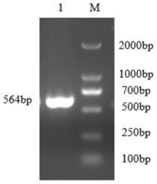
[0072] Embodiment 1 The amplification of PRV gE and gB gene
[0073] 1. Experimental operation
[0074] The upstream and downstream primers were designed respectively according to the whole genome sequence of herpes virus KU056477.1 and the whole genome sequence of herpes type 1 Guangdong isolate with ID KT948041.1 provided by NCBI GenBank (see Table 1). Since it is impossible to design suitable primers for cloning the full-length gene directly using DNA as a template, two sets of primers were designed for two rounds of cloning to obtain PRV gE and gB truncated genes.
[0075] Table 1 Primers:
[0076]
[0077]
[0078] In the first round of amplification, gEF1, gER1, gBF1, and gBR1 were used to amplify PRV gE and gB genes, respectively.
[0079] In the first round of amplification, gEF2, gER2, gBF2, and gBR2 were used to amplify PRV gE and gB genes, respectively. The second round of primers contains EcoR I and HindШ restriction sites, and the bold content in the tabl...
Embodiment 2
[0083] Construction and identification of embodiment 2 recombinant plasmids pMAL-c5x PRV gE, pMAL-c5x PRV gB
[0084] 1. Experimental operation
[0085] The obtained PRV gE and PRV gB gene fragments were respectively ligated with the pMAL-c5X expression vector after double digestion with EcoR I and HindШ restriction endonucleases, and named as pMAL-c5x PRV gE and pMAL-c5x PRV gB respectively. After the ligation product was transformed, the single colony picked was subjected to the identification of the recombinant plasmid after expansion: (1) PCR; (2) double digestion of the two recombinant plasmids with restriction endonucleases EcoR I and HindШ; ( 3) Send the extracted plasmid to Sangon Biotechnology Co., Ltd. for sequencing.
[0086] 2. Experimental results
[0087] After PCR, gel electrophoresis obtained specific bands of about 546bp and 609bp, respectively, which were consistent with the expected size.
[0088] After double enzyme digestion, gel electrophoresis, pMAL-c...
Embodiment 3
[0090] Example 3 Detection of antigen induced expression and soluble analysis, Western-blot verification
[0091] 1. Experimental operation
[0092] (1) Induced expression of antigen
[0093] The positive recombinant plasmids successfully sequenced were transformed into E. coli BL21(DE3) Escherichia coli, and the inducer isopropylthiogalactopyranoside (IPTG) was added at a final concentration of 0.3mM, and the expression was induced in a shaker at 16°C for 8h.
[0094] (2) Solubility analysis of antigen
[0095] Centrifuge the induced bacterial solution at 8000rpm / min for 10min, discard the supernatant, and resuspend the bacterial cells with PBS. The resuspended cells were ultrasonically disrupted in an ice bath, 200W, working for 3 sec, intermittent for 5 sec, until the bacterial liquid became clear. The bacterial solution was centrifuged at 12000rpm at 4°C for 20min, the supernatant and the precipitate were collected respectively, and the precipitate was resuspended in PBS ...
PUM
| Property | Measurement | Unit |
|---|---|---|
| Sensitivity | aaaaa | aaaaa |
| Sensitivity | aaaaa | aaaaa |
Abstract
Description
Claims
Application Information
 - R&D
- Intellectual Property
- Life Sciences
- Materials
- Tech Scout
- Unparalleled Data Quality
- Higher Quality Content
- 60% Fewer Hallucinations
Browse by: Latest US Patents, China's latest patents, Technical Efficacy Thesaurus, Application Domain, Technology Topic, Popular Technical Reports.
© 2025 PatSnap. All rights reserved.Legal|Privacy policy|Modern Slavery Act Transparency Statement|Sitemap|About US| Contact US: help@patsnap.com


