Magnetic-particle separation chemiluminescence immunoassay for detecting glial fibrillary acidic protein (GFAP)

A technology of chemiluminescence immunity and glial fiber, which is applied in the direction of chemiluminescence/bioluminescence, and analysis by making materials react chemically, and can solve the problems of low automation of ELISA method, large pollution of RIA method, complicated and cumbersome operation, etc.

Inactive Publication Date: 2019-03-26
BEIJING LEADMAN BIOCHEM
View PDF8 Cites 15 Cited by
  • Summary
  • Abstract
  • Description
  • Claims
  • Application Information

AI Technical Summary

Problems solved by technology

However, there are certain defects in these methods. The RIA method is highly polluted, the ELISA method is low in automation, and the operation is complicated and cumbersome. High performance liquid chromatography, gas chromatography, and gas chromatography and mass spectrometry are not suitable for widespread clinical testing.

Method used


Image

  • Magnetic-particle separation chemiluminescence immunoassay for detecting glial fibrillary acidic protein (GFAP)
  • Magnetic-particle separation chemiluminescence immunoassay for detecting glial fibrillary acidic protein (GFAP)
  • Magnetic-particle separation chemiluminescence immunoassay for detecting glial fibrillary acidic protein (GFAP)

Examples


Embodiment 1

[0047] Example 1: Magnetic particle-labeled glial fibrillary acidic protein (GFAP) antibody

[0048] Take 20 mg of carboxyl-modified microparticle solution, settle the magnetic particles with superparamagnetic properties, uniform particle size, and carboxyl (COOH-) active groups on the surface (magnetic separation) under the action of a magnetic field for 10 minutes, remove the supernatant, and settle the magnetic particles Wash 3 times with (2-(N-morpholine)ethanesulfonic acid) MES at a molar concentration of 0.05M in the activation buffer, pH 6.0 buffer solution, 2 ml each time.

[0049] After washing, the magnetic particles were fully suspended with 1.0ml activation buffer (2-(N-morpholine) ethanesulfonic acid) MES with a molar concentration of 0.05M, pH 6.0, and then the activator 1-ethyl-3- [3-dimethylaminopropyl]carbodiimide hydrochloride (EDC), suspension reaction at room temperature for 30 minutes, the molar concentration of EDC is 7.5mM.

[0050] Take 1.0 mg of glial...

Embodiment 2

[0052] Example 2: Alkaline phosphatase (ALP) labeled glial fibrillary acidic protein (GFAP) antibody

[0053] Take 1.0 mg of glial fibrillary acidic protein (GFAP) antibody, concentrate to 2.5 mg / mL, add 5 μL of activator 2-Iminothiolane hydrochloride (2-IT) solution with a concentration of 13.76 mg / mL, and react at room temperature for 15 minutes. Use Sephadex G25 gel column to desalt and collect the activated antibody.

[0054] Take 1.2 mg of alkaline phosphatase (ALP), concentrate it to 2.5 mg / mL, add 12 μL of activator Succinimidyl 4-(N-maleimidomethyl)cyclohexane-1-carboxylate (SMCC) solution with a concentration of 6.69 mg / mL, and react at room temperature for 15 minute. Use Sephadex G25 gel column to desalt and collect activated alkaline phosphatase (ALP).

[0055] Mix activated glial fibrillary acidic protein (GFAP) antibody and alkaline phosphatase (ALP) according to the ratio of 1.0 mg glial fibrillary acidic protein (GFAP) antibody to 1.0 mg alkaline phosphatase (...

Embodiment 3

[0057] Embodiment 3: Human Tau protein (TAU) magnetic particle separation chemiluminescent immunoassay method

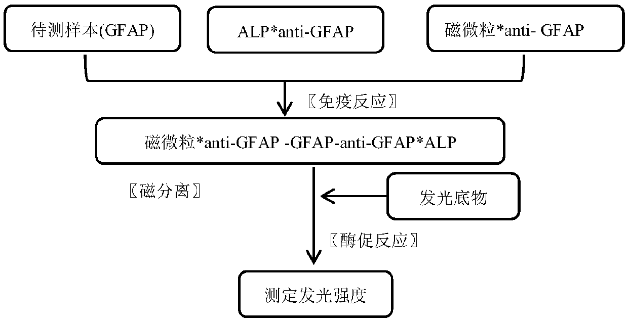
[0058] (1) Immunological reaction: the 50 μl calibrator series of Example 1 (concentrations are respectively 0, 0.8, 2.5, 9.5, 25, 50 ng / ml) were respectively mixed with the quality control substance, 50 μl reagent A of Example 1, Example 1 Add 50 μl of reagent B of 2 to the reaction tube in turn, mix and incubate at 37°C for 15 minutes;

[0059] (2) Magnetic separation: settle the magnetic particles in a magnetic field, remove the supernatant, add 200-500 μl of cleaning solution, remove the magnetic field, then settle the magnetic particles in the magnetic field again, and remove the supernatant; repeat this 2-4 times, To remove unbound antibodies and impurities;

[0060] (3) Reading value: add 150 μl of luminescence substrate solution, after alkaline phosphatase (ALP) catalyzes the luminescence of the substrate, measure the relative luminescence intensity (RLU) wi...


PUM


Abstract

The invention discloses a magnetic-particle separation chemiluminescence immunodetection method for glial fibrillary acidic protein (GFAP) of a human body. A kit comprises a calibration product, a quality control product reagent A, a reagent B, concentrated cleaning liquid and luminescent substrate liquid, wherein the calibration product is antigen containing a series concentrations of glial fibrillary acidic proteins and is used for establishing standard curves; the quality control product is prepared by buffering solution of antigens containing certain-concentration glial fibrillary acidic protein; the reagent A is antibody solution containing certain-concentration glial fibrillary acidic protein and labeled by magnetic particles; the reagent B is antibody solution containing certain-concentration glial fibrillary acidic protein and labeled by the alkaline phosphatase; the concentrated cleaning liquid is used for preparing cleaning liquid; the luminescent substrate liquid is luminescent substrate solution catalyzed by alkaline phosphatase (ALP). The magnetic-particle separation chemiluminescence immunodetection method disclosed by the invention has the beneficial effects that thesignal intensity and the sensitivity of immunoreaction are greatly improved, low-content substances also can generate strong chemiluminescence signals when in immunobinding, and a method with more accuracy, precision, convenience and simpleness is provided for detecting the glial fibrillary acidic protein of the human body.

Description

technical field [0001] The invention belongs to the technical field of immunoassay and analysis, and provides a magnetic separation chemiluminescence immunoassay method for detecting glial fibrillary acidic protein (GFAP), which is suitable for the quantitative detection of human serum glial fibrillary acidic protein (GFAP). Background technique [0002] Glial fibrillary acidic protein (GFAP) is a specific acidic protein in the cytoplasm of astrocytes (AS) in the central nervous system. In recent years, many studies have found that serum and cerebrospinal fluid GFAP levels have important guiding significance in the diagnosis, treatment and prognosis of neurological diseases, which has attracted more and more attention. [0003] 1. Overview of GFAP [0004] The human GFAP gene is located in zone 2, zone 2, long arm of chromosome 17 (17q21), has 8 introns and 9 exons, and has high homology with the coding regions of mouse and rat genes. GFAP was originally isolated from matu...

Claims


Application Information

Patent Timeline
Patent Type & Authority Applications(China)
IPC IPC(8): G01N21/76
CPCG01N21/76
Inventor 郭健夫桂颖袁方
Owner BEIJING LEADMAN BIOCHEM
Why Eureka
Try Eureka