Stent for directional induction of neural stem cell differentiation and preparation method thereof

A neural stem cell and directional induction technology, which is applied in the field of scaffolds for directional induction of neural stem cell differentiation and its preparation, can solve the problems of non-degradable and low efficiency of silica gel sheets, and achieve the goal of promoting adhesion growth, good application prospects, and restoring functionality Effect

Inactive Publication Date: 2019-06-14
SHANDONG BRANDEN MEDICAL DEVICE
View PDF10 Cites 4 Cited by
  • Summary
  • Abstract
  • Description
  • Claims
  • Application Information

AI Technical Summary

Problems solved by technology

[0004] Patent CN 102181396 B discloses a method for inducing the directional differentiation of neural stem cells into sensory neurons in vitro. Stem cells are planted on a silica gel membrane, and the development and maturation of sensory neurons are induced by tension traction stimulation. The sensory neurons differentiated by this method Neurons account for 5% of the total number of neural stem cell differentiation, the efficiency is low, and the silica gel sheet used is non-degradable, requiring a second operation to remove it

Method used


Image

  • Stent for directional induction of neural stem cell differentiation and preparation method thereof
  • Stent for directional induction of neural stem cell differentiation and preparation method thereof
  • Stent for directional induction of neural stem cell differentiation and preparation method thereof

Examples


Embodiment 1

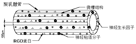
[0050] 1) Mix PLGA-(ASP-PEG)n-PLGA copolymer, 20 μg nerve growth factor and 20 μg nerve adhesion molecule

[0051] Add to 50ml PLGA solution; ultrasonically mix, and form a catheter by electrospinning, forming conditions: positive voltage 10kv, negative voltage 1.5kv;

[0052] 2) Using a femtosecond laser to engrave microgrooves with a groove width of 100 μm on the inner and outer walls of the catheter prepared in step 1), a groove depth of 10 μm, and a groove spacing of 50 μm, and sterilize by irradiation;

[0053] 3) Put the sterilized scaffold tube with microgrooves in step 2) into 5ml of neural stem cell solution, and then culture it in a 37°C, 5% CO2 incubator, induce culture for 14 days, and replace it every three days culture medium to observe cell proliferation and differentiation;

[0054] The results showed that the adhesion rate of the cells was 95%, and about 85% of the neural stem cells with an aspect ratio of about 5 grew along the orientation of the microgroove...

Embodiment 2

[0056] 1) Add PLGA-(ASP-PEG)n-PLGA copolymer, 20μg nerve growth factor and 20μg fibronectin to 50ml PLGA solution; ultrasonically mix, and form a catheter by electrospinning, forming conditions: positive voltage 10kv , the negative voltage is 1.5kv;

[0057] 2) Using a femtosecond laser to engrave microgrooves with a groove width of 100 μm on the inner and outer walls of the catheter prepared in step 1), a groove depth of 10 μm, and a groove spacing of 50 μm, and sterilize by irradiation;

[0058] 3) Put the sterilized scaffold tube with microgrooves in step 2) into 5ml of neural stem cell solution, and then culture it in a 37°C, 5% CO2 incubator, induce culture for 14 days, and replace it every three days culture medium to observe cell proliferation and differentiation;

[0059] The results showed that about 80% of neural stem cells with an aspect ratio of about 5 grew along the microgroove orientation.

Embodiment 3

[0061] 1) Add PLGA-(ASP-PEG)n-PLGA copolymer, 20μg nerve growth factor and 10μg fibronectin to 50ml PLGA solution; ultrasonically mix, and form a catheter by electrospinning, forming conditions: positive voltage 10kv , the negative voltage is 1.5kv;

[0062] 2) Using a femtosecond laser to engrave microgrooves with a groove width of 100 μm on the inner and outer walls of the catheter prepared in step 1), a groove depth of 10 μm, and a groove spacing of 50 μm, and sterilize by irradiation;

[0063] 3) Put the sterilized scaffold tube with microgrooves in step 2) into 5ml of neural stem cell solution, and then culture it in a 37°C, 5% CO2 incubator, induce culture for 14 days, and replace it every three days culture medium to observe cell proliferation and differentiation;

[0064] The results showed that about 75% of neural stem cells with an aspect ratio of about 5 grew along the microgroove orientation.


PUM

PropertyMeasurementUnit
thicknessaaaaaaaaaa
diameteraaaaaaaaaa
diameteraaaaaaaaaa

Abstract

The invention discloses a stent for directional induction of neural stem cell differentiation and a preparation method thereof, and belongs to the field of polymer materials and biomedical engineering. The adhesion growth of cells is promoted by improving the hydrophilicity of a matrix material of the stent; a microgroove structure is constructed on the inner and outer surfaces of the stent material, and the function of directional induction of the differentiation is achieved; at the same time, the stent can further release biological active factors in the controlled mode and promote the adhesion and the growth of the stem cells after the directional differentiation. The stent has a good application prospect in neural stem cell transplantation.

Description

technical field [0001] The invention belongs to the fields of polymer materials and biomedical engineering, and in particular relates to a scaffold for directional induction of neural stem cell differentiation and a preparation method thereof. Background technique [0002] Peripheral nerve injury has a high morbidity rate, with more than 5 million cases of PNI occurring each year worldwide. Acute trauma-induced peripheral nerve injury (PNI) can lead to complete loss of motor function and muscle paralysis, with the radial nerve in the upper extremity and the peroneal nerve in the lower extremity being the most commonly injured nerves. Peripheral nerves typically repair damaged nerves by attempting to regenerate, yet traumatic injury to peripheral nerves remains a daunting clinical challenge. For long-gap nerve repair, autologous nerve transplantation is still the best clinical choice, but autologous transplantation often leads to loss of function of the donor nerve site. In...

Claims


Application Information

Patent Timeline
Patent Type & Authority Applications(China)
IPC IPC(8): A61L27/18A61L27/22A61L27/54A61L27/58A61F2/02
Inventor 张海军曹文瑞鲁手涛杨飞张明广康全利刘黎明周超尹玉霞侯文博段翠海刘光
Owner SHANDONG BRANDEN MEDICAL DEVICE
Why Eureka
Try Eureka