
Preparation method of graphene-coated atomic force microscope probe

An atomic force microscope and graphene coating technology, which is applied in scanning probe technology, scanning probe microscopy, measuring devices, etc., can solve the problems of many impurities in the coating of the tip of the needle, inability to large-scale preparation, and high production costs, and achieve The effect of reducing electrical conductivity and mechanical properties, reducing the amount of oxidation, and avoiding pollution

Inactive Publication Date: 2019-11-29
YANCHENG TEACHERS UNIV
View PDF0 Cites 1 Cited by
  • Summary
  • Abstract
  • Description
  • Claims
  • Application Information

AI Technical Summary

Problems solved by technology

However, the current related preparation methods, such as impregnation method, transfer method, etc., still have the shortcomings of too many impurities coated on the needle tip, which cannot be prepared on a large scale, the process is very complicated, and the production cost is high.

Method used


Image

  • Preparation method of graphene-coated atomic force microscope probe
  • Preparation method of graphene-coated atomic force microscope probe
  • Preparation method of graphene-coated atomic force microscope probe

Examples


Embodiment 1

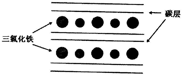
[0030] (1) Mix 300mg of anhydrous ferric chloride and 60mg of expanded graphite evenly, vacuumize, seal in a 50mL glass bottle, and heat at 400°C for 4h to prepare a pure second-order graphite intercalation compound. Dissolve the graphite intercalation compound in Dilute hydrochloric acid solution, filter and dry.

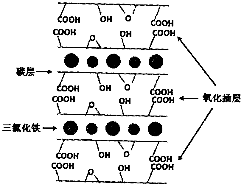
[0031] (2) The graphite intercalation compound was added into a mixed solution of 20 mL of concentrated sulfuric acid and 10 mL of concentrated nitric acid, stirred in ice water (0° C.) for 0.5 hour, 360 mg of sodium chlorate was put into the solution, and stirred at room temperature for 12 hours, The product was washed by centrifugation.

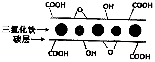
[0032] (3) The product is added to an excess of hydrogen peroxide with a mass fraction of 30%, and the reaction time is 1 h to obtain single-sided selective graphene oxide.

[0033] (4) Put the single-sided selective hydrophilic graphene material into water, and the single-sided selective hydrophilic graphene material is disper...

Embodiment 2

[0038] (1) Mix 300mg of anhydrous ferric chloride and 60mg of expanded graphite evenly, vacuumize, seal in a 50mL glass bottle, and heat at 400°C for 4h to prepare a pure second-order graphite intercalation compound. Dissolve the graphite intercalation compound in Dilute hydrochloric acid solution, filter and dry.

[0039] (2) The graphite intercalation compound was added into a mixed solution of 20 mL of concentrated sulfuric acid and 10 mL of concentrated nitric acid, stirred in ice water (0° C.) for 0.5 hour, 360 mg of sodium chlorate was put into the solution, and stirred at room temperature for 12 hours, The product was washed by centrifugation.

[0040] (3) The product is added to an excess of hydrogen peroxide with a mass fraction of 30%, and the reaction time is 1 h to obtain single-sided selective graphene oxide.

[0041] (4) Put the single-sided selective hydrophilic graphene material into water, and the single-sided selective hydrophilic graphene material is disper...


PUM


Abstract

The invention provides a preparation method of a graphene-coated atomic force microscope probe. The preparation method comprises the following steps of preparing a single-sided selective graphene oxide material, and preparing a single-layer graphene film on a gas-liquid interface in a self-assembly manner; placing the atomic force microscope probe to be coated below the liquid level, and enablingthe atomic force microscope probe to be at a positive potential; slowly pumping the liquid to descend the liquid level, and covering the tip of the atomic force microscope probe with the single-layergraphene; and implementing microwave treatment, so that a metal layer can effectively absorb microwave energy, and internal oxygen-containing functional groups are decomposed. According to the method,the single-layer graphene film is prepared at the gas-liquid interface in the self-assembly manner, the film can be controlled at the thickness of a monatomic layer, graphene is firmly attached to the surface of the atomic force microscope probe, coating is tight and uniform, the service life of the probe is prolonged, the preparation process is simple, used materials are easy to obtain, the requirement for equipment is low, large-scale and batch coating can be achieved, and the production efficiency is greatly improved.

Description

technical field [0001] The invention relates to the field of atomic force microscopy, in particular to a method for preparing a graphene-coated atomic force microscopy probe. Background technique [0002] Atomic force microscope (AFM) is an important instrument for the analysis of surface topography and electromagnetic properties with atomic resolution. Atomic force microscope probes (AFM probes) are generally composed of silicon (Si) or silicon nitride (Si 3 N 4 ), the tip radius of the probe is generally 10 to tens of nm, and the microcantilever is usually a silicon wafer or a silicon nitride wafer with a length of 100-500 μm, a width of 10-50 μm, and a thickness of 500 nm-5 μm. A conductive AFM probe is obtained by plating 10-50nm thick metal platinum (and other metals that improve the bonding force of the coating, such as titanium, chromium, platinum and iridium, etc.) on the surface of the common probe. However, the conductive coating of conductive AFM probes is easy...

Claims


Application Information

Patent Timeline
IPC IPC(8): G01Q60/38G01Q70/16
CPCG01Q60/38G01Q70/16
Inventor 苗中正张立云
Owner YANCHENG TEACHERS UNIV
Learn More