A preparation method of multilayer spiral urethral tissue engineering scaffold

A tissue engineering scaffold and urethral technology, which is applied in the field of preparation of tissue engineered urethral scaffolds for urethral diseases, can solve the problems of sudden disintegration, hinder the physiological activity of the urethra, and become brittle, and achieve the effects of avoiding blockage of the urethra and reducing the surface coverage.

Active Publication Date: 2021-07-27
SOUTHEAST UNIV
View PDF1 Cites 0 Cited by
  • Summary
  • Abstract
  • Description
  • Claims
  • Application Information

AI Technical Summary

Problems solved by technology

[0003] The defect of the existing urethral stent preparation method is that although the stent formed by curling the polymer film is easy to manufacture and has high compliance, it is prone to sudden disintegration during the degradation process, forming larger fragments, which is easy to cause The urethra is blocked, which hinders the normal physiological activities of the urethra; in addition, relying solely on the self-expandability of the spiral stent, the expansion rate is relatively slow, and there is a possibility of dislocation after stent implantation
As a result, the printed product is basically in a glass state at room temperature, with low strength and high brittleness, which cannot meet the mechanical performance requirements of the bracket.
[0006] Simply implanting the carrying cells into the vicinity of the defect in the organism is prone to the problem of cell death due to insufficient supply of nutrients

Method used


Image

  • A preparation method of multilayer spiral urethral tissue engineering scaffold
  • A preparation method of multilayer spiral urethral tissue engineering scaffold
  • A preparation method of multilayer spiral urethral tissue engineering scaffold

Examples


Embodiment 1

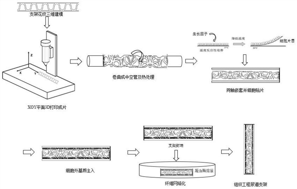
[0043] A kind of preparation of urethra tissue engineering support, comprises the steps:

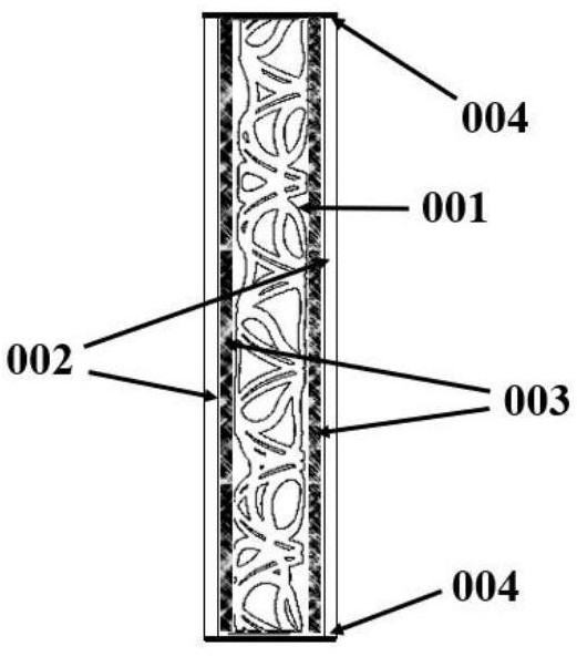
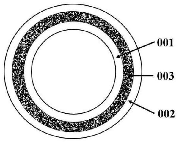
[0044] (1) Using polylactic acid-glycolic acid (PLGA) copolymer as raw material (LA:GA=75:25, Mv=100000), after three-dimensional modeling, import the .stl file into the fused deposition 3D printer and print out two surfaces A rectangular long embryo body with a coverage rate of 20% and a spline curve pattern; the wall thickness of the support, that is, the thickness of the rectangular long embryo body is 0.1 mm; 1 3.0mm and φ 2 On a 5.6mm mold; both ends are fixed with thin copper wires; then placed in a vacuum oven at 120 ° C for 10 minutes to promote polymer crystallization and the shape of the bracket; after cooling, take out the mold to obtain monolayers with different diameters The bracket is then cut into a long tube with a length of 30.0 mm.

[0045] (2) The stents of equal length and matching diameter are coaxially nested and fixed to form a double-layer stent, and one end of ...

Embodiment 2

[0054] A kind of preparation of urethra tissue engineering support, comprises the steps:

[0055] (1) Using polylactic acid (molecular weight: 100,000) as the raw material, import the .stl file into the fused deposition 3D printer after 3D modeling and print out two rectangular strips with a surface coverage of 30% and spline curve patterns Body, the wall thickness of the stent, that is, the thickness of the rectangular long strip embryo body is 0.3mm; respectively, two rectangular long strip embryo bodies are helically crimped with diameters of φ 1 = 4.0mm and φ 2 = 6.8mm on the mold; both ends are respectively fixed with thin copper wires; then placed in a vacuum oven at 90°C for 10 minutes to be annealed and shaped; after the mold is cooled, the mold is taken out to obtain single-layer stents of different diameters, which are all cut to length A long tube of 40.0mm.

[0056] (2) A double-layered stent is formed by coaxially nesting and fixing stents of equal length and ma...

Embodiment 3

[0065] A kind of preparation of urethra tissue engineering support, comprises the steps:

[0066] (1) Using polycaprolactone (molecular weight: 50,000) as the raw material, import the .st1 file into the fused deposition 3D printer after three-dimensional modeling and print out two rectangles with a surface coverage of 40% and spline curve patterns. Strip-shaped embryo body, the wall thickness of the support, that is, the thickness of the rectangular long strip embryo body is 0.4mm; two rectangular long strip embryo bodies are helically crimped on diameters of φ 1 = 4.5mm and φ 2 = 7.3mm on the mold; both ends are fixed with thin copper wires; then placed in a vacuum oven at 30°C for annealing for 20 minutes and finalized; after the mold is cooled, the mold is taken out to obtain single-layer brackets with different diameters, which are all cut to length A long tube of 60.0mm.

[0067](2) The stents of equal length and matching diameter are coaxially nested and fixed to form ...


PUM

PropertyMeasurementUnit
thicknessaaaaaaaaaa
thicknessaaaaaaaaaa
thicknessaaaaaaaaaa

Abstract

A method for preparing a multilayer spiral urethral tissue engineering scaffold, using thermoplastic degradable polymers as printing materials, using a fused deposition 3D printer to prepare a rectangular long strip embryo body with a hollow pattern, and spirally curling to obtain a spiral tube of the embryo body , after heat treatment and nesting, a double-layer scaffold composed of inner and outer embryoid body spiral tubes was obtained, and one end of the double-layer scaffold was sutured with polylactic acid sutures, and transfected when cultured sequentially on the inner wall of the outer embryoid body spiral tube Endothelial cell sheets of vascular endothelial growth factor and fibroblast growth factor, urothelial cell sheets, and smooth muscle cell sheets transfected with human platelet growth factor during culture, filled with extracellular matrix, and then sutured into the double-layer scaffold On the other end, the scaffold for urethral tissue engineering can be obtained after being fixed with thrombin. The stent prepared by the invention can keep the urethra unobstructed, is beneficial to the healing of the damaged urethral tissue, realizes the vascularization of the urethral stent in time, and can quickly realize blood supply reconstruction.

Description

technical field [0001] The invention belongs to the field of biomedical devices, and in particular relates to a preparation technology for a tissue-engineered urethral stent for treating urethral diseases such as urethral defects. [0002] Background of the invention [0003] The defect of the existing urethral stent preparation method is that although the stent formed by curling the polymer film is easy to manufacture and has high compliance, it is prone to sudden disintegration during the degradation process, forming larger fragments, which is easy to cause Urethral blockage hinders normal physiological activities of the urethra; in addition, relying solely on the self-expandability of the helical stent, the expansion rate is relatively slow, and there is a possibility of dislocation after stent implantation. The preparation process of the braided stent is slow, inefficient, time-consuming and lacks precise control of the stent structure; the compliance is insufficient and ...

Claims


Application Information

Patent Timeline
Patent Type & Authority Patents(China)
IPC IPC(8): C12N5/07A61L31/12A61L31/14A61L31/16A61L31/00B29C64/118B33Y80/00
CPCA61L31/005A61L31/129A61L31/14A61L31/148A61L31/16A61L2300/30A61L2300/604B33Y80/00B29C64/118
Inventor 白晶王先丽张磊薛烽程兆俊范启洲
Owner SOUTHEAST UNIV
Why Eureka
Try Eureka