Microcatheter with releasable coating at head end

A micro-catheter and coating technology, applied in the direction of catheter and coating, can solve the problems of safety risks in the catheter, and achieve the effect of excellent biological safety, avoiding potential risks and reducing the withdrawal force.

Active Publication Date: 2020-01-17
SUZHOU HENGRUI HONGYUAN MEDICAL TECH CO LTD
View PDF12 Cites 2 Cited by
  • Summary
  • Abstract
  • Description
  • Claims
  • Application Information

AI Technical Summary

Problems solved by technology

[0007] However, the shortcoming of the above three designs is that they will leave a microcatheter tip about 5cm long in the body, and the catheter trapped in the body may still pose a safety risk

Method used


Image

  • Microcatheter with releasable coating at head end
  • Microcatheter with releasable coating at head end
  • Microcatheter with releasable coating at head end

Examples


Embodiment 1

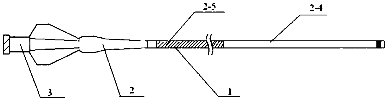
[0036] Such as Figure 1-2 As shown, this embodiment provides a microcatheter, including a microcatheter main body 1, a stress expansion tube 2 and a Luer connector 3; the microcatheter main body 1 is connected to one end of the stress expansion tube 2, and the other end of the stress expansion tube 2 It is connected with the Luer connector 3; the surface of the microcatheter main body 1 is coated with a detachable double coating.

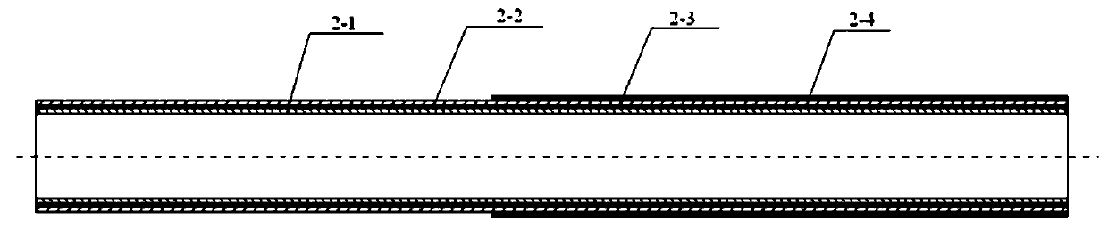
[0037] The cross-sectional structure of the microcatheter main body 1 includes an inner layer 2-1, an intermediate layer 2-2 and an outer layer 2-3 from the inside to the outside; the material of the inner layer 2-1 is a PTFE catheter, and the middle layer 2-2 It is a metal lining wire layer of braided or coiled spring structure, and the material of the outer layer 2-3 is PEBAX catheter.

[0038] The PEBAX catheter in the outer layer 2-3 is welded by several sections of PEBAX materials with different hardness; wherein the distal end material is so...

Embodiment 2

[0041] Weigh 14g of polyvinylpyrrolidone, 36g of acrylic acid, 35.5g of acrylamide, dissolve in water, add 0.01g of N,N'-methylenebisacrylamide and 0.01g of ammonium persulfate, and react at 70°C under nitrogen protection 3h, immerse the 5cm tip of the microcatheter described in Example 1 in the solution for 5s, then lift it up and away from the liquid surface at a speed of 20mm / s, place the microcatheter in an oven, and dry it at 30°C.

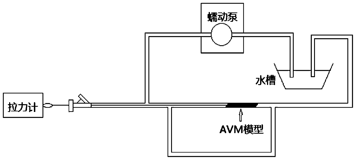
[0042] Use Onyx glue to test the withdrawal force of the microcatheter, press image 3 Build the test pipeline, the test method is as follows:

[0043] (1) Soak the dipped microcatheter in 37°C normal saline for 10 minutes;

[0044] (2) Build a microcatheter in vitro detection model as shown in the figure

[0045] (3) The water flow rate in the pipeline is 40mL / min

[0046] (4) Insert the microcatheter into the arteriovenous malformation model and make close contact with it

[0047] (5) Slowly inject the liquid embolism into the arteriove...

Embodiment 3

[0058] Weigh polyethylene glycol 10g, chitosan 10g, dissolve in deionized water containing 2% acetic acid, add a certain amount of 2 × 10⁻⁵mol / ml glutaraldehyde aqueous solution, filter and let stand for degassing, and the 1. The tip 7cm of the microcatheter is dipped in the solution for 5s, then lifted up and away from the liquid surface at a speed of 20mm / s, and the microcatheter is placed in an oven and dried at 30°C.

[0059] Using the test method described in Example 2, the measured withdrawal force of the microcatheter is 15±7g.


PUM


Abstract

The invention discloses a microcatheter with a releasable coating at the head end. The head end of the microcatheter is coated with the release-type coating, and the release-type coating is of a single-layer structure. The invention further discloses that the release-type coating is a semi-interpenetrating network polymer coating. The invention further discloses a method for preparing the head-endelutable semi-interpenetrating network coating. The semi-interpenetrating network polymer coating does not swell or swell slowly in water, but can quickly swell in a dimethyl sulfoxide solvent of a liquid embolic agent, so that a polymer I in the release-type coating is released, the adhesion of a micelle formed by solidifying the liquid embolic agent to the microcatheter I is reduced, the microcatheter is withdrawn as a whole, damage to the blood vessel due to inability to withdraw the microcatheter or excessive force of microcatheter withdrawal is avoided, the microcatheter head-end is notleft in a body, and risk of microcatheter withdrawal is effectively reduced.

Description

technical field [0001] The invention relates to a medical microcatheter, in particular to a detachable microcatheter with a double-coated head end, which belongs to the technical field of medical instruments. Background technique [0002] Cerebral arteriovenous malformation (AVM) is a congenital local cerebrovascular variation disease. Intracranial blood vessels lose their normal vascular tissue structure and lack capillary beds between arteries and veins, resulting in abnormal arteriovenous shunts and venous hypertension. Risk of rupture. [0003] The existing treatment methods for AVM mainly include surgical resection, radiotherapy, and endovascular embolization. Endovascular embolization can seal AVM lesions, reduce AVM volume, block aneurysms and fistulas, and improve the safety of surgery and radiotherapy. [0004] Among them, the application of new liquid embolic agents (OnyxTM, PhilTM, etc.) through intravascular embolization is an important method for the treatment...

Claims


Application Information

Patent Timeline
Patent Type & Authority Applications(China)
IPC IPC(8): A61L29/12A61L29/04A61L29/06A61L29/08A61L29/02A61M25/00
CPCA61L29/02A61L29/041A61L29/06A61L29/085A61M25/0045A61M2025/0047C08L27/18
Inventor 刘建民李强吴可沁解荡
Owner SUZHOU HENGRUI HONGYUAN MEDICAL TECH CO LTD
Why Eureka
Try Eureka