
Artificial structure for rapidly regenerating vascularized tissue as well as construction method and application of artificial structure

A construction method and vascularization technology, applied in the field of artificial structures and their construction, can solve the problems of insufficient nutrient supply, cell necrosis, slow growth of blood vessels into implants, etc.

Active Publication Date: 2020-05-22
TSINGHUA UNIV
View PDF6 Cites 3 Cited by
  • Summary
  • Abstract
  • Description
  • Claims
  • Application Information

AI Technical Summary

Problems solved by technology

[0004] Another major challenge for the survival of large-volume implants in vivo is rapid and sufficient angiogenesis, because when the distance between each cell and the nearest blood vessel is greater than 200 μm, there is a problem of insufficient nutrient supply, which eventually leads to cell necrosis.
In the absence of external conditions, the growth rate of blood vessels into the implant is extremely slow, generally on the order of a few millimeters per day

Method used


Image

  • Artificial structure for rapidly regenerating vascularized tissue as well as construction method and application of artificial structure
  • Artificial structure for rapidly regenerating vascularized tissue as well as construction method and application of artificial structure
  • Artificial structure for rapidly regenerating vascularized tissue as well as construction method and application of artificial structure

Examples


Embodiment 1

[0143] Example 1 Preparation of islet cells / hydrogel microspheres

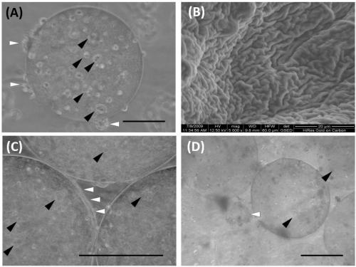
[0144] Take conventionally cultured human pancreatic cells, use a sodium alginate / collagen mixture as a carrier material, and use a high-voltage electrostatic spray preparation device to make pancreatic cell / hydrogel microspheres:

[0145] 1. A solution of sodium alginate (Sigma-Aldrich, A0682) and a collagen solution (Sigma, C7661) were mixed to form carrier material solutions with final concentrations of 2% (w / w) and 1 mg / mL (w / v), respectively.

[0146] 2. Human pancreatic cells (PriCells, HUM-CELL-0058) were mixed with 10 6 The density per ml is uniformly mixed with the carrier material solution and filled into a disposable syringe with a volume of 5 ml.

[0147] 3. Connect the high-voltage electrostatic spraying cell microsphere preparation device. The high-voltage electrostatic generating device adopts SA167-Y (Tianjin) high-voltage electric field generator with an output voltage of 10kV; clamp the syri...

Embodiment 2

[0154] Example 2 Preparation of hepatic parenchymal cells / hydrogel microspheres

[0155] Take conventionally cultured human hepatic parenchymal cells and use sodium alginate / gelatin system as the carrier material, and use a high-voltage electrostatic spraying cell microsphere preparation device to make hepatocyte / hydrogel microspheres:

[0156] 1. Mix a solution of sodium alginate (Sigma-Aldrich, A0682) and a solution of gelatin (Sigma-Aldrich, G1890) (solvent PBS buffer solution or saline) to a final concentration of 2% (w / w) and 3% respectively (w / w) carrier material solution.

[0157] 2. Hepatocytes (PriCells, HUM-CELL-0036) were mixed with 10 6 The density per ml is uniformly mixed with the carrier material solution and filled into a disposable syringe with a volume of 10 ml.

[0158] 3. Connect the high-voltage electrostatic spraying cell microsphere preparation device. The high-voltage electrostatic generating device adopts SA167-Y (Tianjin) high-voltage electric field...

Embodiment 3

[0167] Example 3 Preparation of cervical cancer tumor cells / hydrogel microspheres

[0168] Take conventionally cultured cervical cancer tumors, prepare human cervical cancer tumor cell / hydrogel microspheres, and use sodium alginate / vitronectin system as the microsphere material:

[0169] 1. Sodium alginate solution and vitronectin solution were mixed to obtain solutions with final concentrations of 2% and 1 mg / ml, respectively, as the microsphere material precursor solution.

[0170] 2. Cervical cancer cells (ATCC, H1HeLa) were treated with 10 7 The density of each / ml is uniformly mixed with the carrier material precursor solution and filled into a disposable syringe with a volume of 10ml.

[0171] 3. Referring to step 3 in Example 2, cervical cancer tumor cell / sodium alginate / vitronectin microspheres were prepared.

[0172] 4. Collect the cell microspheres within 5 minutes, wash twice with normal saline and observe and record. The shape of the cell microspheres is round and...


PUM

PropertyMeasurementUnit
The average diameteraaaaaaaaaa

Abstract

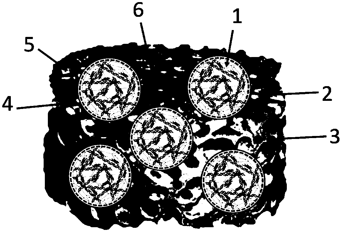
The invention provides an artificial structure capable of being used for rapidly regenerating vascularized tissue and a construction method of the artificial structure. The artificial structure includes cell / hydrogel microspheres, tissue-related cells, vascularized cells, growth factor slow-release microspheres and a carrier material, wherein tissue-related cells are uniformly distributed in the cell / hydrogel microspheres, and the outsides of the microspheres contain a vascularized cell layer; and the cell / hydrogel microspheres, the tissue-related cells, the vascularized cells and the growth factor slow-release microspheres are uniformly distributed in the carrier material. The artificial structure provided by the invention can be cultured in a bioreactor to obtain a vascularized tissue precursor with a vascular-like network structure, or transplanted into the body to regenerate vascularized functional tissue. The obtained tissue precursor or functional tissue has certain physiologicalfunctions and can be used for in-vivo and in-vitro studies such as tissue development, disease occurrence, drug development and detection, tissue regeneration and repair.

Description

technical field [0001] The invention relates to the field of biotechnology, in particular to an artificial structure that can be used to rapidly regenerate vascularized tissue and its construction method and application. Background technique [0002] In the 1980s, tissue engineering began to be applied to research in the medical field, with the goal of developing and eventually applying biological substitutes to restore, maintain or improve tissue function. Through tissue engineering, bioengineered tissues can be designed and prepared in the laboratory. The engineered tissue can be implanted into the body to restore, replace, maintain or improve the function of tissues and organs. [0003] In vivo transplantation of engineered tissue can be divided into two types: traumatic transplantation and minimally invasive surgery. In clinical use, engineered tissue must have a large volume of centimeters and encapsulate a large number of cells to complete the repair or replacement o...

Claims


Application Information

Patent Timeline
IPC IPC(8): C12N5/071A61L27/52A61L27/50A61L27/58A61L27/38
CPCA61L27/3808A61L27/3834A61L27/3895A61L27/507A61L27/52A61L27/58C12N5/0691C12N2501/11C12N2501/115C12N2501/135C12N2501/148C12N2501/165C12N2531/00
Inventor 姚睿孙伟
Owner TSINGHUA UNIV
Learn More