Exosome preparation method and application

A technology of exosomes and detection methods, applied in the biological field, can solve the problems of not being able to preserve the patient's in situ tissue, not being able to provide intercellular, tissue cells and extracellular matrix information, and achieve early diagnosis, signal strength and retention Time stabilization, the effect of increasing the accuracy of quantification

Pending Publication Date: 2020-07-21
CENT FOR EXCELLENCE IN MOLECULAR CELL SCI CHINESE ACAD OF SCI
View PDF4 Cites 0 Cited by
  • Summary
  • Abstract
  • Description
  • Claims
  • Application Information

AI Technical Summary

Problems solved by technology

In existing studies, exosomes are mostly derived from cell culture medium or serum, plasma and other body fluids, which cannot retain the characteristics of the patient's in situ tissue, and cannot provide information on cells, tissue cells and extracellular matrix

Method used


Image

  • Exosome preparation method and application
  • Exosome preparation method and application
  • Exosome preparation method and application

Examples


Embodiment 1

[0070] Example 1 Preparation of exosomes

[0071] The organoid culture supernatant was provided by the cooperative hospital Changhai Hospital. The specific preparation method of the organoid culture supernatant was as follows: the tissue samples from patients with pancreatic cancer were chopped into fine pieces with surgical scissors, and placed in tissue digestion solution ( Human complete medium including 1 mg / mL Collagenase II and 10.5 μM Y-27632) in medium temperature and shaking for 1 hour, filter out undigested tissue with a 70 μm filter membrane, count the cell suspension, and plant according to 1 well of each 6-well plate The density of 200,000 cells was planted, and the resulting cells were planted on the artificial basement membrane (Matrigel) and cultured in human complete feeding medium (Humancomplete Feeding Medium, the formula is: advanced DMEM / F12, HEPES10mM, Glutamax 1X, A83-01500nM, mNoggin 100ng / mL, hFGF10 100ng / mL, hGastrin I 0.01μM, N-acetylcysteine1.25mM, ...

Embodiment 2

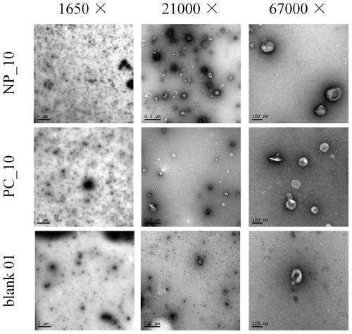
[0079] Embodiment 2 transmission electron microscope detection

[0080] 1) Take 25 μL of exosome sample and drop it on the copper grid, incubate at room temperature for 1 minute, and then use filter paper to dry the solution along the edge of the copper grid;

[0081] 2) Then add 25 μL of 2% uranyl acetate staining solution dropwise, incubate at room temperature for 1 minute, and absorb the solution along the edge of the copper grid with filter paper;

[0082] 3) Put it under the lamp and bake for 10 minutes. After the surface of the copper mesh is dried, observe it with a transmission electron microscope, and record the field of view at magnifications of 1700 times, 21000 times, and 67000 times, respectively.

Embodiment 3

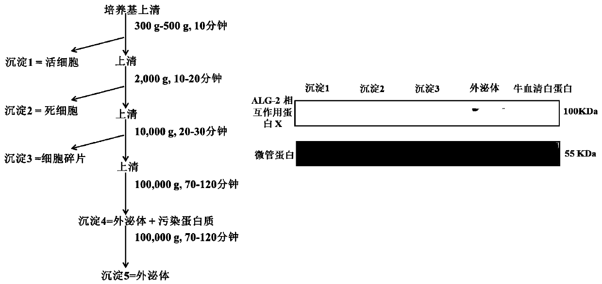
[0083] Example 3 Western blot detection of exosomes

[0084] Each pellet obtained by differential centrifugation was freeze-dried, and 20 μL of SDS lysate (4% SDS, 100 mM Tris, 0.1 MDTT, pH 7.6) was added. At the same time, configure a negative control, add 20 μL SDS lysate to 20 μg BSA. Add 5 μL of 5×sample buffer (0.25M Tris, 10% SDS, 50% glycerol, 0.5% bromophenol blue), and centrifuge in boiling water for 10 minutes to prepare the sample. Then follow the steps below:

[0085] 1) Configure 6% upper glue and 12% lower glue, 15mA constant current, 2.5 hours;

[0086] 2) Transfer to membrane, wet transfer protein onto nitrocellulose membrane (PVDF), 100 constant voltage, 1.5 minutes;

[0087] 3) Put the membrane into the blocking solution (5% BSA, H2O) at room temperature for 1 hour;

[0088] 4) Incubate the primary antibody and incubate overnight at 4 degrees;

[0089] 5) After incubating the primary antibody, wash the membrane 5 times with TBST solution (150mM NaCl, 20m...


PUM

PropertyMeasurementUnit
diameteraaaaaaaaaa

Abstract

The invention relates to an exosome preparation method and application. The method comprises at least the following steps: a culture supernatant of an organoid is processed via a differential centrifugation method, and an exosome is obtained after separation. According to the preparation method, the exosome can be separated and purified in a high-efficiency way; the exosome of the organoid can reserve in-situ tissue characteristics and can provide intercellular, histiocytic and extracellular matrix information; and the exosome of the organoid can provide better targeted and more comprehensivein-situ tissue features and is beneficial to the research on inter-cancer-cellular matrix and cancer cellular and cytoplasmic matrix and to the advancement of cancer treatment.

Description

technical field [0001] The invention belongs to the field of biotechnology, and in particular relates to a preparation method and application of exosomes. Background technique [0002] Exosomes are secreted by most cells, with a diameter of about 30-150 nanometers, tiny membrane vesicles with a lipid bilayer membrane structure, which contain specific proteins, lipids and nucleic acids. Exosomes not only play a role in important physiological activities such as stem cell differentiation and maintenance, tissue repair and tissue regeneration, immune system, embryo implantation and placental development in pregnancy, and reproduction, but also play a role in pathological processes such as cardiovascular diseases, neurodegenerative diseases, and cancer. Activities also play a role. [0003] Exosomes play an important role in the process of tumor proliferation and metastasis. In cancer, exosomes mainly have three functions: regulating tumor immune response, regulating tumor mic...

Claims


Application Information

Patent Timeline
Patent Type & Authority Applications(China)
IPC IPC(8): C12N5/09C12N5/071C12Q1/02G01N33/68G01N33/574
CPCC12N5/0693C12N5/0676G01N33/5011G01N33/6848G01N33/57438C12N2503/02G01N2500/10
Inventor 曾嵘吴家睿李辰陈曦
Owner CENT FOR EXCELLENCE IN MOLECULAR CELL SCI CHINESE ACAD OF SCI
Why Eureka
Try Eureka