
Method for coating mycoplasma pneumoniae membrane protein antigens with magnetic beads

A technology of Mycoplasma pneumoniae and membrane protein, applied in the biological field, can solve the problems of low efficiency of Mycoplasma pneumoniae membrane protein antigen coating magnetic beads, complex protein components, etc., and achieve the effect of improving coating efficiency and stability

Pending Publication Date: 2020-08-25
ZHUHAI LIVZON DIAGNOSTICS
View PDF9 Cites 5 Cited by
  • Summary
  • Abstract
  • Description
  • Claims
  • Application Information

AI Technical Summary

Problems solved by technology

[0005] CN110672837A discloses a kind of technological method of magnetic bead coating protein, what this method adopts is carboxyl magnetic bead, applicable coating protein is TSH antibody, AFP antibody and TG antigen, does not include the mycoplasma pneumoniae membrane protein antigen, and this method aims at Improve the coating efficiency of the protein, and the stability of the protein after the magnetic bead coating is not involved
[0006] CN110045116A discloses a coating accelerator that can effectively improve the coating efficiency of viral antigens, but the method is mainly to add reducing agents, protein denaturants, nonionic surfactants and organic reducing agents during the antigen coating process to release antigens Antibody binding sites to improve the coating efficiency of purified viral antigens, this method is aimed at recombinant viral antigens, non-viral antigens, especially the application of natural Mycoplasma pneumoniae membrane protein antigens is not mentioned, and the coating carrier of this method is micropores plates instead of beads
[0007] The protein composition of the Mycoplasma pneumoniae membrane protein antigen extracted from the naturally cultured Mycoplasma pneumoniae is complex, so that the efficiency of the Mycoplasma pneumoniae membrane protein antigen coated magnetic beads is very low

Method used


Image

  • Method for coating mycoplasma pneumoniae membrane protein antigens with magnetic beads
  • Method for coating mycoplasma pneumoniae membrane protein antigens with magnetic beads
  • Method for coating mycoplasma pneumoniae membrane protein antigens with magnetic beads

Examples


Embodiment 1

[0078] (a) Prepare 1M dithrethiol, pH7.4 phosphate buffer, 50M CB buffer, and 1M ammonium sulfate for later use.

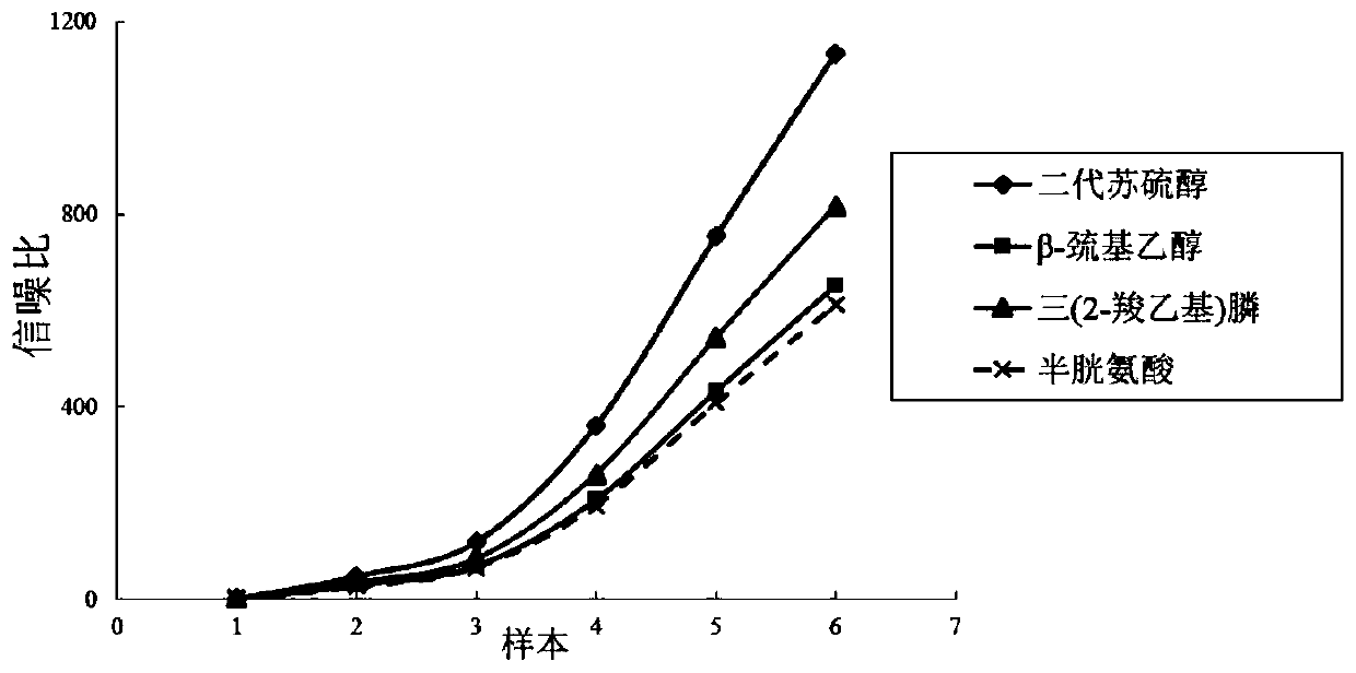
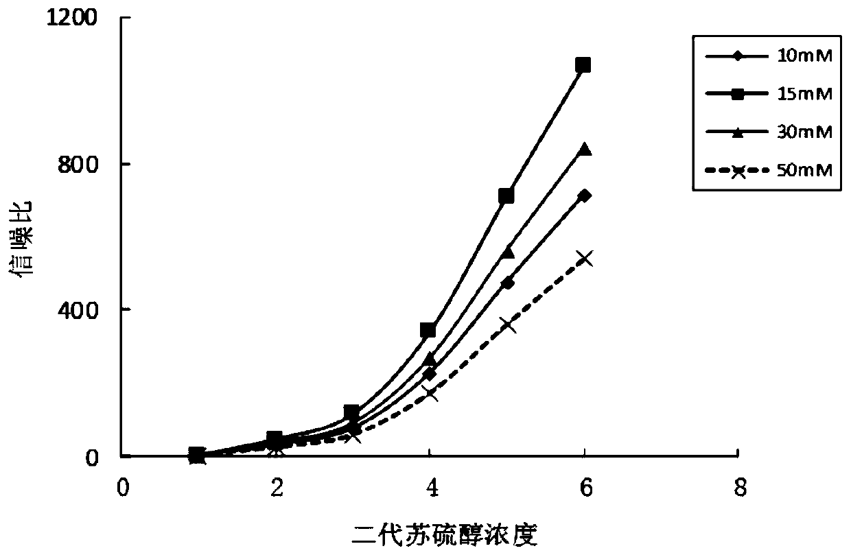
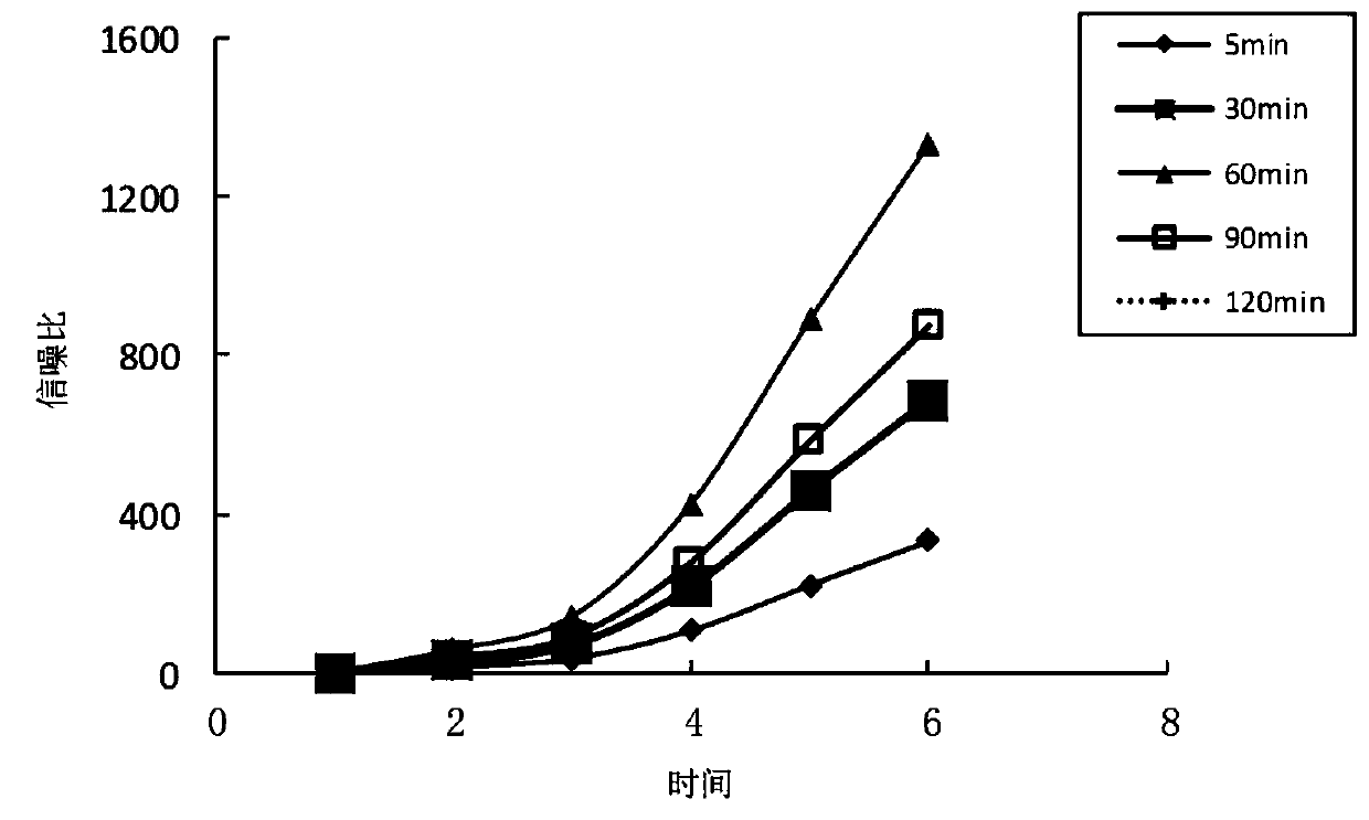
[0079] (b) Second-generation threothiol was added to the Mp membrane protein antigen to make a final concentration of 15 mM, placed in an environment at 4°C, and rotated on a rotary mixer at a speed of 20 rpm for 60 min to make it evenly mixed.

[0080] (c) Put the above solution into the treated dialysis bag, tie the mouth of the bag well and put it into a beaker filled with pH7.4 phosphate buffer for dialysis, replace with new buffer every 3 hours, repeat 3 times.

[0081] (d) Wash the Tosyl magnetic beads three times with 1 mL of phosphate buffer, and discard the supernatant by magnetic suction.

[0082] (e) Add 200 μL of CB buffer, Mp membrane protein antigen (15 μg / mg Tosyl magnetic beads) and 100 μL of ammonium sulfate, and place it on a rotary mixer at 20 rpm for 2 hours at 37 ° C to make the antigen and Magnetic bead coupling.

[0083] (f) Wash the magne...

Embodiment 2

[0088] The method for coating magnetic beads with Mp membrane protein antigen provided in this example differs from Example 1 only in that the reducing agent is tris(2-carboxyethyl)phosphine.

Embodiment 3

[0090] The method for coating magnetic beads with Mp membrane protein antigen provided in this example differs from Example 1 only in that the reducing agent is β-mercaptoethanol.


PUM


Abstract

The invention provides a method for coating mycoplasma pneumoniae membrane protein antigens with magnetic beads, and relates to the technical field of biology. The mycoplasma pneumoniae membrane protein antigens coated by the method are natural mycoplasma pneumoniae protein. The method comprises the following steps: treating the mycoplasma pneumoniae membrane protein antigens by using a reducing agent, and coupling the mycoplasma pneumoniae membrane protein antigens with magnetic beads. According to the method, the technical problem that the efficiency of coating the magnetic beads with the natural mycoplasma pneumoniae membrane protein antigen is low is alleviated.

Description

technical field [0001] The invention relates to the field of biotechnology, in particular to a method for coating magnetic beads with mycoplasma pneumoniae membrane protein antigen. Background technique [0002] Mycoplasma pneumonia (Mp) is a tiny prokaryotic pathogenic microorganism that lacks a cell wall and is naturally resistant to antibacterial drugs that act on the cell wall (such as penicillins and cephalosporins). It is a common pathogen of respiratory tract infections in children. Mp is mainly transmitted by droplets, and the incubation period in the body is 2-3 weeks, and it is infectious from the incubation period to the remission of symptoms for several weeks. Mp infection can also cause changes in various extrapulmonary systems, and severe cases can lead to death. Therefore, timely and effective detection of Mp infection is of great significance. [0003] At present, the diagnosis methods of Mp infection adopted at home and abroad include classical culture met...

Claims


Application Information

Patent Timeline
Patent Type & Authority Applications(China)
IPC IPC(8): G01N33/543G01N33/569
CPCG01N33/54326G01N33/56933G01N2333/30G01N2469/20
Inventor 侯雨晴朱越谭曾敏霞储迅涛
Owner ZHUHAI LIVZON DIAGNOSTICS
Learn More