Bilirubin derivative-based diagnostic and therapeutic ultrasound contrast agent

A technology of bilirubin and derivatives, applied in the field of ultrasound contrast agents, can solve the problem of low image quality

Pending Publication Date: 2020-10-09
株式会社毕丽斯
View PDF7 Cites 3 Cited by
  • Summary
  • Abstract
  • Description
  • Claims
  • Application Information

AI Technical Summary

Problems solved by technology

However, since the image quality is low compared with MRI, CT, and PET techniques, various ultrasound contrast agents are being developed to improve the image quality

Method used


Image

  • Bilirubin derivative-based diagnostic and therapeutic ultrasound contrast agent
  • Bilirubin derivative-based diagnostic and therapeutic ultrasound contrast agent
  • Bilirubin derivative-based diagnostic and therapeutic ultrasound contrast agent

Examples


Embodiment 1

[0093] Embodiment 1: Preparation of the ultrasound contrast agent based on pegylated bilirubin of the present invention

[0094] 1-1. Preparation of bilirubin derivatives (pegylated bilirubin, Pegylated bilirubin)

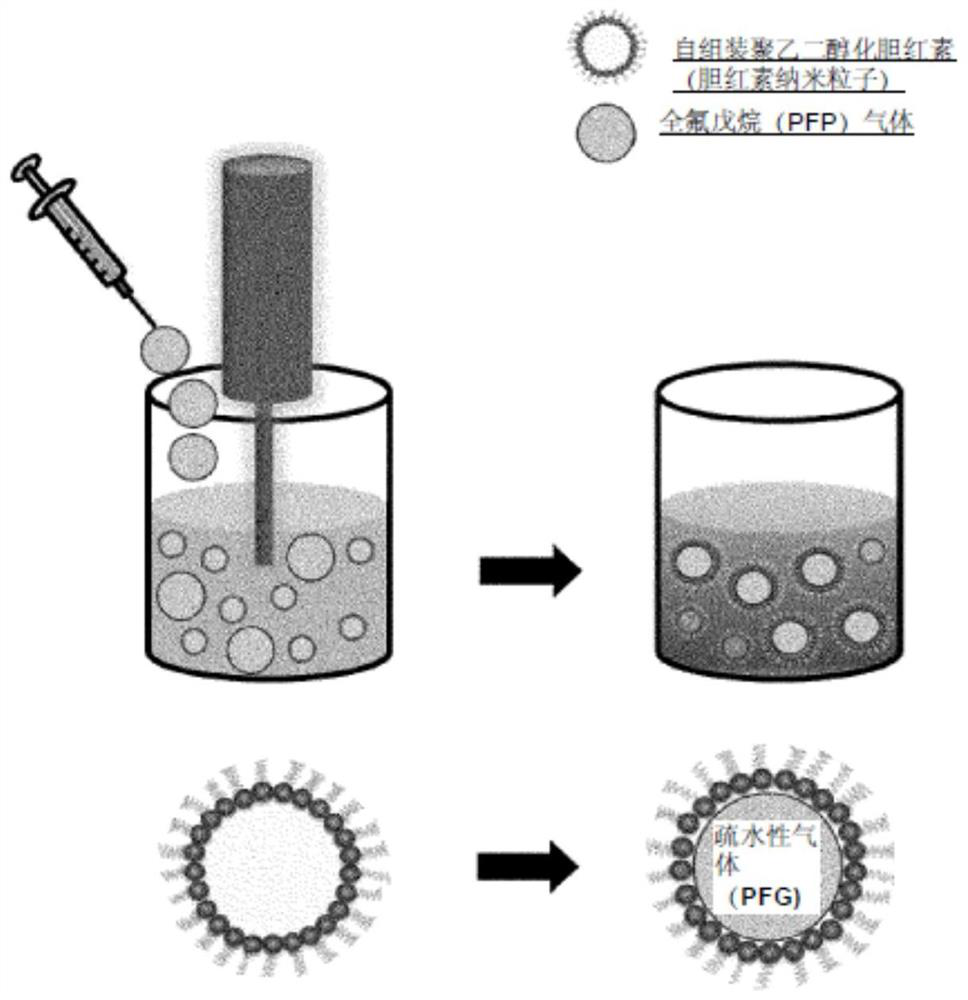
[0095] The present inventors prepared amphiphilic derivatives of bilirubin in which a hydrophilic molecule was bound to bilirubin prior to preparation of a bilirubin-based ultrasound contrast agent. As the hydrophilic molecule, polyethylene glycol (polyethyleneglycol) was used.

[0096] Specifically, first dissolve bilirubin in dimethylsulfoxide (DMSO), and then add an appropriate amount of EDC (1-Ethyl-3-(3-dimethylaminopropyl)carbodiimide) to activate the carboxyl groups present in bilirubin , so as to induce the desired reaction, and react at room temperature for 10 minutes. Thereafter, polyethylene glycol having an amine group at the end is added and reacted for a period of time to synthesize a bilirubin derivative (Pegylated bilirubin) in which the carboxyl ...

Embodiment 2

[0100] Example 2: Phantom Imaging of Pegylated Bilirubin-Based Ultrasound Contrast Agents of the Invention

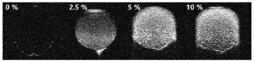
[0101] Ultrasound phantom imaging was acquired using a Vevo770 (High-Resolution Micro-Imaging System, Visualsonics, Toronto, Canada) equipped with an ultrasound device probe for mice, RMV 706 probe. In order to simulate in vivo conditions for ultrasound imaging, the present inventors used an agarose-gel phantom made by embedding 500 μL unit microcentrifuge tubes in 3% (w / v) produced in agarose gel.

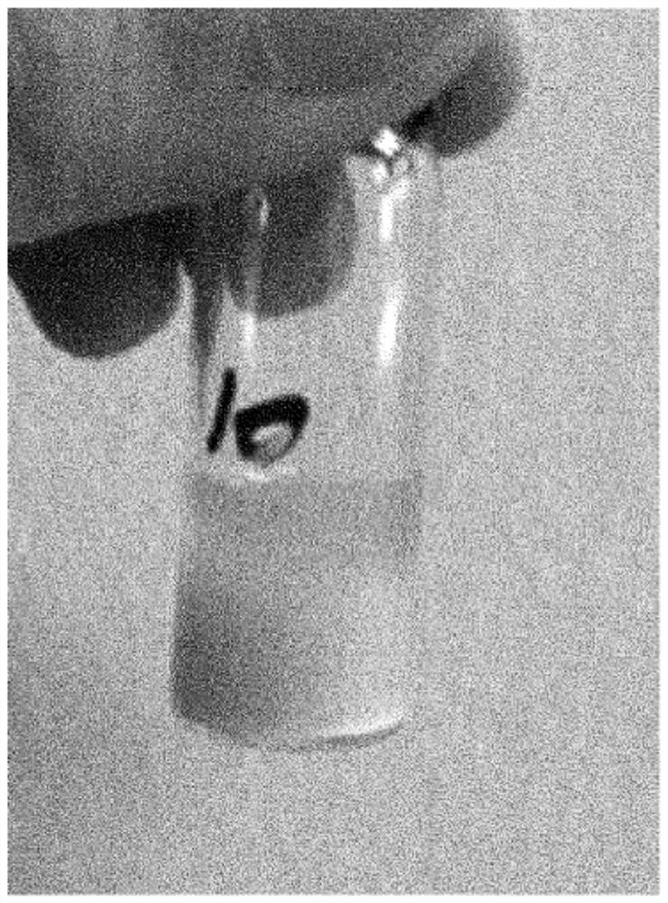
[0102] First, 300 μL of PEGylated bilirubin-based contrast agent samples (PFP ​​0, 2.5, 5, 10% v / v) of the present invention using perfluoropentane (perfluoropentane, PFP) as the hydrophobic gaseous core Placed in an agarose-gel phantom, and images were acquired by 40 MHz ultrasound. The change in ultrasonic intensity of each sample (PFP ​​respectively 0, 2.5, 5, 10% v / v) was measured over a period of 180 minutes, and as a normalization process, the water control group wa...

Embodiment 3

[0106] Example 3: Characteristics of the Pegylated Bilirubin-Based Ultrasound Contrast Agent of the Invention

[0107] 3-1. Microscopic morphology

[0108] Microscopic morphology of fine particles was performed by negative staining with uranium acetate and transmission electron microscopy (Tecnai G2 F30, Eindhoven, Netherlands) ( Figure 6 ) and the optical microscope under the coverslip ( Figure 7 ) to observe.

[0109] Figure 6 This is a picture of a pegylated bilirubin-based ultrasound contrast agent observed with a transmission electron microscope (TEM, transmission electron microscopy). Figure 6 Micron-sized air bubble particles constituting the ultrasound contrast agent of the present invention are shown. Figure 7 is a pegylated bilirubin-based ultrasound contrast agent for observation by light microscopy.

[0110] In addition, in order to calculate the number of air bubbles contained per volume of the contrast agent, the pegylated bilirubin-based contrast agent o...


PUM

PropertyMeasurementUnit
molecular weightaaaaaaaaaa
sizeaaaaaaaaaa

Abstract

The present invention relates to a bilirubin derivative-based diagnostic and therapeutic ultrasound contrast agent. Fine particles comprising a bilirubin derivative according to the present inventionreact sensitively to reactive oxygen species (ROS), bond to a hydrophobic drug and enable effective chelating of metals such as iron oxide nanoparticles. Therefore, the fine particles according to thepresent invention can be utilized as a contrast agent for an ultrasonic diagnosis as well as a contrast agent for a magnetic resonance image diagnosis and a carrier for hydrophobic and platinum-baseddrugs.

Description

technical field [0001] This patent application claims priority to Korean Patent Application No. 10-2018-0014160 filed with the Korean Patent Office on February 5, 2018, and the disclosure of the patent application is incorporated herein by reference. [0002] The invention relates to an ultrasonic contrast agent for diagnosis and treatment based on bilirubin derivatives. Background technique [0003] Ultrasound (ultrasound) refers to a sound wave with a frequency greater than the human audible range, that is, 20,000 Hz. Using this ultrasonic wave, various boundaries such as various organs, bones, muscle tissue, and blood in the body will pass through the diffusion, reflection, absorption and absorption of sound waves. The signal difference generated by scattering is called ultrasonic image (ultrasonography). Ultrasound imaging devices for medical diagnosis, as one of the most widely used diagnostic techniques at present, have outstanding mobility and proximity. Compared wit...

Claims


Application Information

Patent Timeline
Patent Type & Authority Applications(China)
IPC IPC(8): A61K49/22A61K31/409A61K51/04A61K47/69A61K9/16
CPCA61K31/409A61K49/225A61K49/1833A61K49/186A61K41/0028A61K47/6923A61K47/6929A61K31/704A61K31/7068A61K31/136A61K31/337A61K9/0085A61K47/46A61K2300/00A61K49/223A61K51/0451A61K47/6939A61K9/1676A61K33/243A61K9/5031A61K31/282A61K49/221
Inventor 全尚勇李东润李勇贤金镇勇
Owner 株式会社毕丽斯
Why Eureka
Try Eureka