Application of a Fluorescent Conjugated Polymer Nanoprobe in Peripheral Nerve Imaging
A conjugated polymer and nanoprobe technology, which is applied to preparations and pharmaceutical formulations for in vivo experiments, to achieve the effects of simple cleaning method, high biological safety, and strong clinical application value.
- Summary
- Abstract
- Description
- Claims
- Application Information
AI Technical Summary
Problems solved by technology
Method used
Image
Examples
Embodiment 1
[0061] Example 1 Preparation and performance characterization of fluorescent conjugated polymer nanoprobes
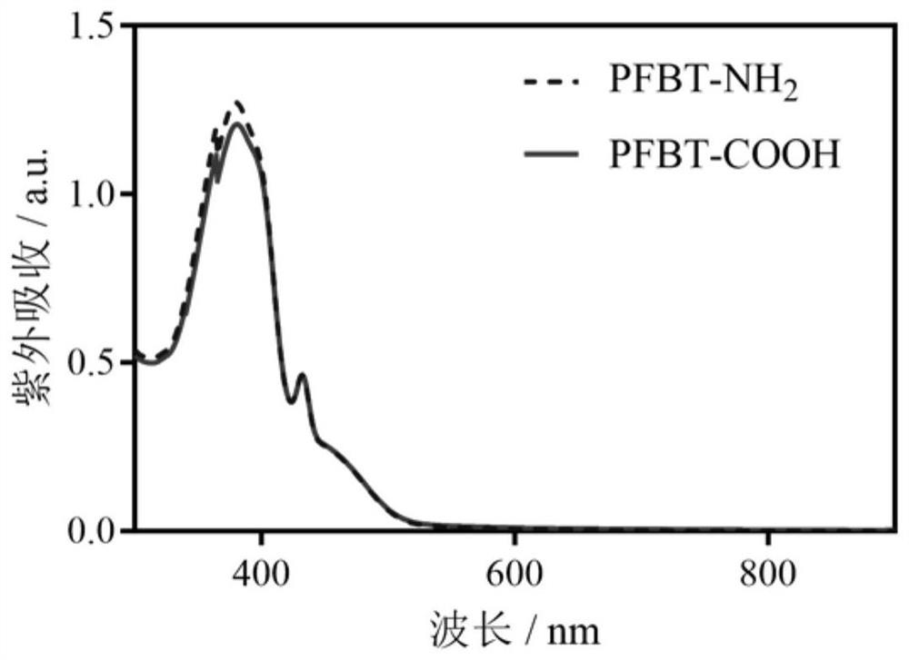
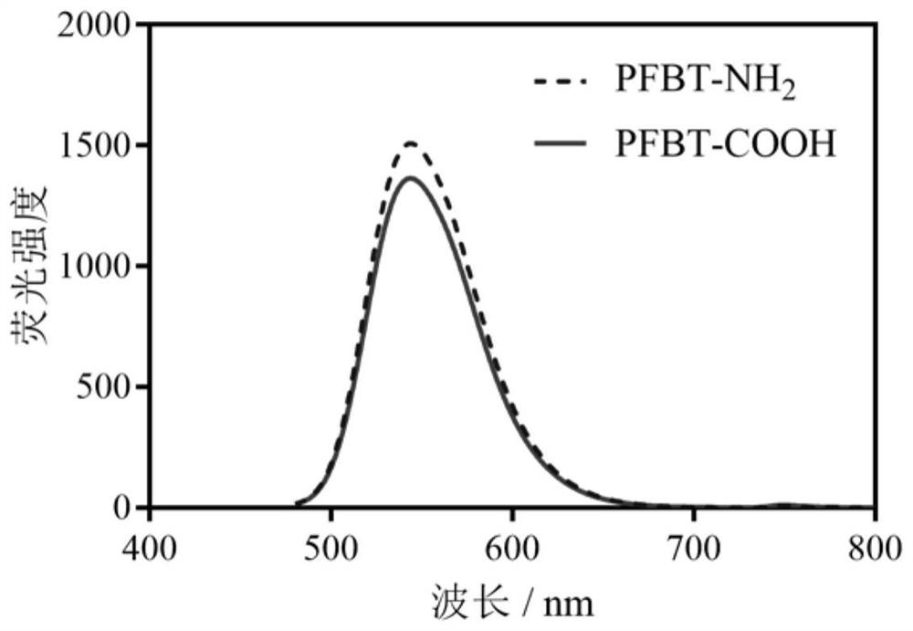
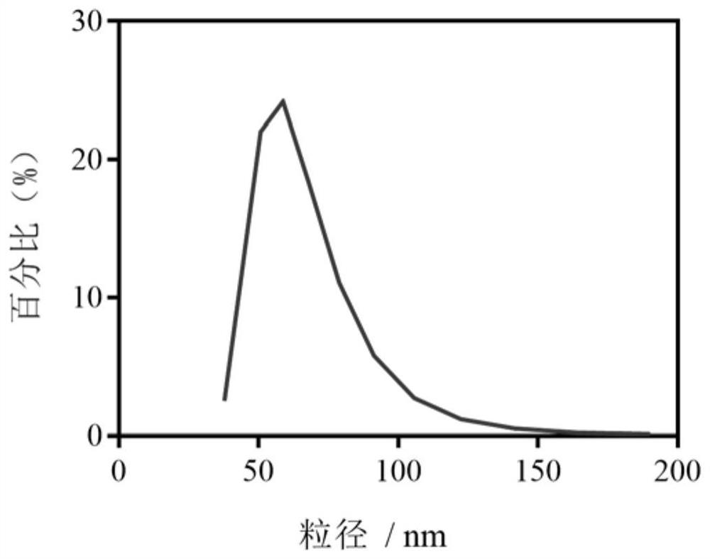
[0062] Take 0.25 mL of poly[(9,9-dioctylfluorenyl-2,7diyl)-(1,4-benzo-{2,1',3}-thiadiazole) at a concentration of 1 mg / mL ] 10% benzothiadiazole (y) (PFBT) solution and 0.25 mL of amino-terminated polymethyl methacrylate (MMA-NH 2 ) solution was added to 1.5mL tetrahydrofuran (THF), in which PFBT stock solution and MMA-NH 2 The storage solution was prepared with THF as solvent. The mixture was sonicated in a water bath for 5 minutes. Under the conditions of probe sonication, the above mixture was quickly transferred to 10 mL of deionized water, and the probe was sonicated for 1 minute. Under the condition of 45℃, N 2 Remove all THF in the solution in 40 minutes to prepare 0.025mg / mL PFBT-NH 2 Fluorescent Conjugated Polymer Nanoprobes.
[0063] Take 0.25 mL of poly[(9,9-dioctylfluorenyl-2,7diyl)-(1,4-benzo-{2,1',3}-thiadiazole) at a concentration of 1 mg / mL ] 10%...
Embodiment 2
[0066] Example 2 Fluorescent Conjugated Polymer Nanoprobes Imaging Mammalian Peripheral Nerves
[0067] The imaging methods of fluorescent conjugated polymer nanoprobes for mammalian peripheral nerves include direct exposure imaging and intramuscular injection imaging, respectively as follows:
[0068] 1. Direct Exposure Imaging
[0069] The mouse sciatic nerve is surgically exposed by removing the cutaneous muscle and fatty tissue overlying the sciatic nerve. 50 μL aqueous solution of fluorescent conjugated polymer nanoprobe was dropped on the exposed left sciatic nerve and right sciatic nerve, covering all tissues (nerve, muscle, fat and fascia) in the surgically exposed area, and the left sciatic nerve was dripped with PFBT -COOH fluorescent conjugated polymer nanoprobe aqueous solution, the right sciatic nerve was dripped with PFBT-NH 2 Fluorescent conjugated polymer nanoprobe aqueous solution, after incubation for a certain period of time, remove each probe aqueous solu...
Embodiment 3
[0075] Example 3 Determination of the binding position of the conjugated polymer nanoprobe with the mouse sciatic nerve
[0076] After the in vivo fluorescence imaging was completed, the mouse sciatic nerve was taken out for the in vitro slice experiment. After embedding in OCT (a water-soluble mixture of polyethylene glycol and polyvinyl alcohol), place it in a -80°C refrigerator for more than 4 hours, cut the tissue into 10 μm thick sections, then perform HE staining, and perform fluorescence on the tissue after sealing. imaging.
[0077] Figure 8 , Figure 9 , Figure 10 Diagram of isolated tissue slices of sciatic nerve in the experimental group, Figure 8 It is the fluorescence imaging and HE staining image of mouse sciatic nerve cross section, Figure 9 It is the fluorescence imaging and HE staining imaging of the longitudinal section of the mouse sciatic nerve. It can be seen from the results of the picture that the probe mainly gathers in the outer nerve model 3 ...
PUM
| Property | Measurement | Unit |
|---|---|---|
| particle size | aaaaa | aaaaa |
Abstract
Description
Claims
Application Information
 - R&D
- Intellectual Property
- Life Sciences
- Materials
- Tech Scout
- Unparalleled Data Quality
- Higher Quality Content
- 60% Fewer Hallucinations
Browse by: Latest US Patents, China's latest patents, Technical Efficacy Thesaurus, Application Domain, Technology Topic, Popular Technical Reports.
© 2025 PatSnap. All rights reserved.Legal|Privacy policy|Modern Slavery Act Transparency Statement|Sitemap|About US| Contact US: help@patsnap.com


