Antibody targeting sars-cov-2 and its preparation method and application

A sars-cov-2, antibody technology, applied in the fields of virology, biomedicine, and immunology, can solve problems such as virus mutation

Active Publication Date: 2021-04-02
SHANGHAI TECH UNIV
View PDF3 Cites 1 Cited by
  • Summary
  • Abstract
  • Description
  • Claims
  • Application Information

AI Technical Summary

Problems solved by technology

However, in the course of future disease treatment, the virus still has the potential to mutate under the selection pressure of antibodies, and this combination strategy does not fundamentally solve the problem of immune escape

Method used


Image

  • Antibody targeting sars-cov-2 and its preparation method and application
  • Antibody targeting sars-cov-2 and its preparation method and application
  • Antibody targeting sars-cov-2 and its preparation method and application

Examples


Embodiment 1

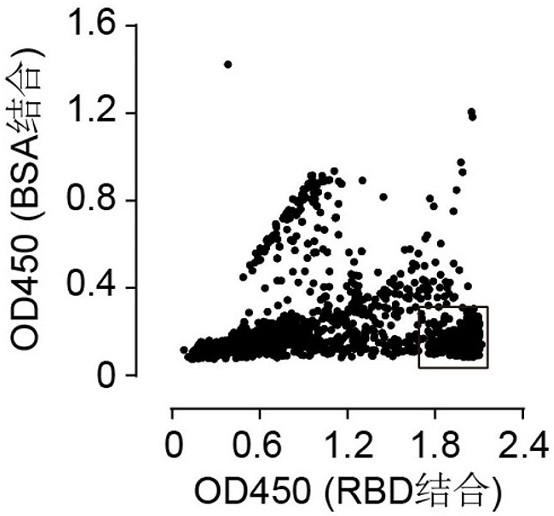
[0078] Example 1. Screening of Human Antibodies Targeting SARS-CoV-2 RBD Using Phage Display Technology

[0079] Referring to previous methods reported (Lim, T. S. et al., V-gene amplification revisited - An optimized procedure for amplification of rearranged human antibody genes of different isotypes. N. Biotechnol. 2010, 27, 108-117), using peripheral blood B cells of recovered patients with new coronavirus, using Trizol (Invitrogen Cat# 15596018) method to extract RNA in B cells, and further using reverse transcription kit (Takara, Cat# R023A) to convert 8 μg of RNA was transcribed into cDNA. The VH gene and VL gene were amplified separately by primers, and finally constructed in the phagemid vector in the form of Fab to obtain a human phage antibody display library. Monoclonal antibodies that specifically recognize and bind to the RBD region of the S protein of the new coronavirus were obtained by panning. The purified RBD protein was coated on a 96-well screening pla...

Embodiment 2

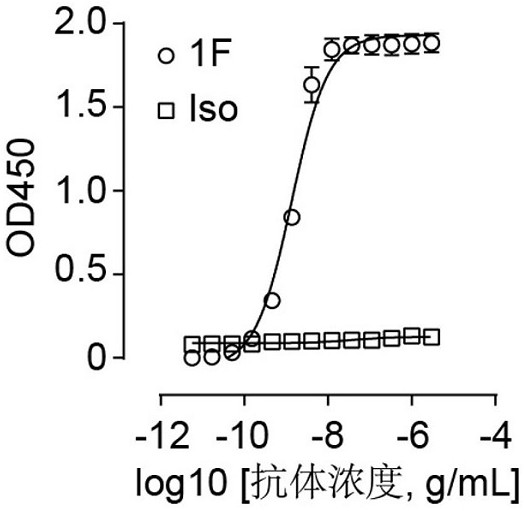
[0084] Example 2. Using ELISA to detect the ability of antibody 1F to bind to RBD

[0085] The immunoassay plate (Thermo Fisher Scientific, Cat# 442404) was coated with 2 μg / mL RBD protein and incubated overnight at 4°C. The next day, the well plate was washed and further blocked with 5% skimmed milk at room temperature for 1 hour. 1F antibody was recombinantly expressed in HEK Expi 293F cells as human IgG1 and purified using protein A Sepharose (GE Healthcare, Cat# 28-9670-56). Add 1F antibodies diluted in different concentrations to the microtiter plate and incubate at room temperature for 1 hour. After washing with PBS, an HRP-conjugated anti-human IgG antibody (Proteintech, Cat#SA00001-17) was added and incubated at room temperature for 1 hour. After washing again, TMB substrate was added to develop color, and after adding 10% sulfuric acid, the absorbance at a wavelength of 450nm was detected. The result is as figure 2 As shown, it can be seen from the figure that an...

Embodiment 3

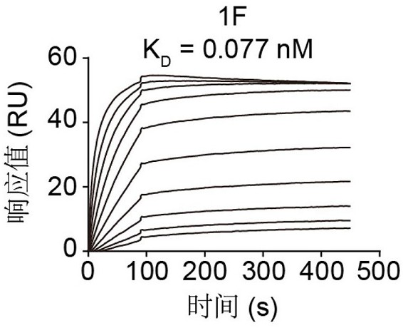
[0086] Example 3. Using Surface Plasmon Resonance (SPR) Technology to Detect the Binding-Dissociation Constant of Antibody 1F and RBD

[0087] The binding-dissociation constant of Antibody 1F was determined using Biacore T200 (GE Healthcare). The purified RBD was immobilized on the surface of the CM5 chip so that the final response unit (RU) was around 100 RU. The SPR test uses PBST solvent with a flow rate of 30 μL / min. The antibody response values ​​at different concentrations were measured, and finally simulated in a 1:1 binding mode to calculate the binding dissociation constant of antibody 1F and RBD. The result is as image 3 shown. K on 1F D The value is 0.077 nM.


PUM


Abstract

The present invention provides an antibody targeting SARS-CoV-2 and its preparation method and application. The antibody comprises VH and VL, and the VH comprises the following CDRs: VH with amino acid sequence as shown in SEQ ID NO: 1, 2, 3 CDR1, VH CDR2, VH CDR3; the VL comprises the following CDRs: VL CDR1, VL CDR2, VL CDR3 with amino acid sequences as shown in SEQ ID NO: 4, 5, 6. The antibody can bind with high affinity and specificity to the RBD of the S protein of SARS-CoV-2, inhibit the binding of the RBD protein to the receptor ACE2 protein, and efficiently inhibit SARS-CoV-2 from infecting cells, while preventing potential immune escape mutations The pseudovirus has good neutralizing activity, so it can be effectively applied to the diagnosis, prevention and treatment of SARS-CoV-2 virus and related diseases.

Description

technical field [0001] The invention relates to the fields of immunology, virology and biomedicine, in particular to an antibody targeting SARS-CoV-2 and its preparation method and application. Background technique [0002] The novel coronavirus SARS-CoV-2 is the pathogen that causes new coronary pneumonia (COVID19), which is highly contagious and spreads widely. SARS-CoV-2 is a single-stranded RNA virus that belongs to betacoronaviruses along with SARS-CoV that causes severe acute respiratory syndrome and MERS-CoV that causes Middle East respiratory syndrome. The SARS-CoV-2 envelope is distributed with a spike protein (spike, S protein), which forms a homotrimer. During the infection process, the S protein can open the receptor binding domain (RBD) and expose The receptor binding motif (RBM) thus binds to the receptor angiotensin converting enzyme 2 (ACE2) on the host cell, and further fuses with the host cell membrane, eventually causing the cell to be infected by the vir...

Claims


Application Information

Patent Timeline
Patent Type & Authority Patents(China)
IPC IPC(8): C07K16/10G01N33/569A61K39/42A61K47/68A61P31/14
CPCA61K2039/505A61P31/14C07K16/10C07K2317/565C07K2319/00G01N33/56983
Inventor 钟桂生王皞鹏赵思蒙邱士真
Owner SHANGHAI TECH UNIV
Why Eureka
Try Eureka