Subunit fusion protein mG on surface of rabies virus as well as preparation method and application of subunit fusion protein mG

A fusion protein and rabies virus technology, applied in biochemical equipment and methods, viruses, viral peptides, etc., can solve the problems of high production cost, inability to produce and apply, low yield, etc., achieve batch-to-batch stability, and facilitate large-scale production , high yield effect

Pending Publication Date: 2021-03-02
NOVO BIOTECH CORP
View PDF1 Cites 1 Cited by
  • Summary
  • Abstract
  • Description
  • Claims
  • Application Information

AI Technical Summary

Problems solved by technology

Although these studies have verified that G protein is a good protective antigen, its yield is very low, and the cost of large-scale industrial production is too high to produce and apply

Method used


Image

  • Subunit fusion protein mG on surface of rabies virus as well as preparation method and application of subunit fusion protein mG
  • Subunit fusion protein mG on surface of rabies virus as well as preparation method and application of subunit fusion protein mG
  • Subunit fusion protein mG on surface of rabies virus as well as preparation method and application of subunit fusion protein mG

Examples


Embodiment 1

[0043] Embodiment 1G protein expression and design

[0044] 1.1 Selection of rabies virus G protein

[0045] The surface envelope protein G protein of rabies virus is a homotrimer in structure, which is an important antigen that induces the body to produce protective neutralizing antibodies. There were in-depth research reports as early as the 1980s and 1990s. Researchers tried to use various The system expresses G protein, but the protein has not been expressed and purified on a large scale. This may be caused by the unstable structure of G protein expressed alone. In order to solve this important technical problem, the present invention introduces a segment at the amino terminus of G protein. To ensure the correct structural folding of the G protein and protein stability, and to improve expression and facilitate screening in CHO cells, we optimized the codons of the encoded mG fusion protein.

[0046] 1.2 Rabies virus G protein codon optimization

[0047] Using the standar...

Embodiment 2

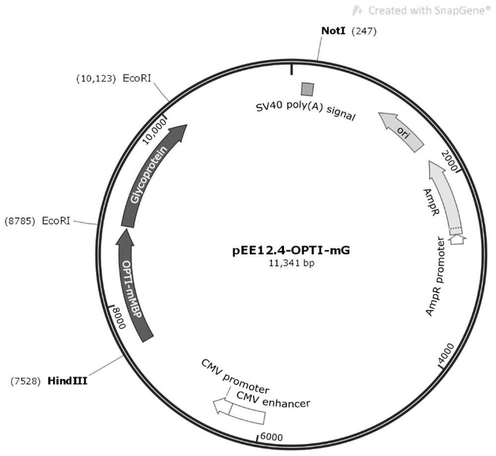
[0048] Example 2: Construction of pEE12.4-OPTI-mG recombinant plasmid

[0049] 2.1PCR amplification of the target fragment OPTI-mG

[0050] 2.1.1 PCR reaction

[0051] (1) Primer design and synthesis

[0052] Upstream primer: 5'-ACGAAGCTTAAGACAGAGGAGGGCAAGC-3'

[0053] Downstream primer: 5'-GAATTGAATTCTCAGTTGGGCAGGCCCAGATC-3'

[0054] (2) Add 50 μL of the sample system, as shown in the table below:

[0055]

[0056] PCR amplification program:

[0057]

[0058] 2.1.2 Gel recovery of PCR products

[0059] (1) Mark the sample collection EP tube, adsorption column and collection tube;

[0060] (2) Take the weight of the marked empty EP tube, and record the value;

[0061] (3) Carefully cut out a single target DNA band from the agarose gel with a scalpel on a gel cutter and put it into a clean 1.5mL centrifuge tube;

[0062] (4) Add 600 μL PC buffer to the 1.5mL centrifuge tube in step (3), place in a 50°C water bath for about 5 minutes, and gently turn the centrifuge...

Embodiment 3

[0109] Example 3: Establishment of transfection of pEE12.4-OPTI-mG recombinant plasmid into CHO-K1 cells and monoclonal screening

[0110] 3.1 CHO-K1 cell transfection

[0111] (1) Preparation: UV sterilization in a biological safety cabinet for 30 minutes; DMEM / F12 (containing 10% serum, 1% double antibody), DMEM / F12 and PBS were placed in a 37°C water bath and preheated to 37°C.

[0112] (2) Take out the cells (10 cm cell culture dish) from the incubator at 37° C., discard the supernatant medium, wash the cells once with pre-warmed 8 mL PBS, and discard the PBS.

[0113] (3) Add 1-2mL 0.25% trypsin-EDTA to each 10cm cell culture dish, digest at room temperature for about 2 minutes, observe under the microscope that the cells shrink and become round, and appear as single cells.

[0114] (4) Add 4 mL of DMEM / F12 (containing 10% serum, 1% double antibody) to terminate the digestion reaction, and blow the cells away with a pipette.

[0115] (5) Transfer the digested cells to a...


PUM

PropertyMeasurementUnit
Molecular weightaaaaaaaaaa

Abstract

The invention provides a subunit fusion protein mG on the surface of rabies virus as well as a preparation method and application of the subunit fusion protein mG. The preparation method comprises thesteps: 1) cloning a gene sequence shown as SEQ ID NO.4 into an eukaryotic expression vector to obtain a recombinant plasmid containing a fusion protein MBP-G encoding gene; 2) transfecting the recombinant plasmid containing the fusion protein MBP-G encoding gene into an expression cell; 3) culturing, screening and domesticating the expression cell in the step 2) to obtain highly expressed cell strains; and 4) fermenting and culturing the cell strain in the step 3), and purifying to obtain the subunit fusion protein mG. MG can be expressed in a large amount in a soluble mode, protein is stable, the problems that rabies virus surface G protein cannot be expressed on a large scale and the like in the prior art are solved, and the preparation method is simple and low in cost.

Description

technical field [0001] The invention belongs to the technical field of recombinant protein expression and purification of biological products, and relates to a subunit fusion protein mG on the surface of rabies virus and its preparation method and application. Background technique [0002] Rabies is a zoonotic infectious disease caused by Rabies virus (RV), with a mortality rate as high as 100%. The clinical manifestations are hydrophobia, photophobia, mania and other symptoms. Dogs are the main host and source of infection for rabies. Rabies is distributed worldwide. The number of deaths caused by rabies is about 40,000 to 70,000 in the world every year, but most of the annual rabies occurs in developing countries, 98% of which are in Asia. my country is the country with the second highest annual rabies incidence in the world after India. Rabies has become an important epidemic that endangers the public health security of our country. [0003] Rabies virus is a member ...

Claims


Application Information

Patent Timeline
IPC IPC(8): C07K19/00C12N15/62C12N15/85G01N33/68G01N33/569A61K39/205A61P31/14
CPCC07K14/005C12N9/1088C12Y205/01018C07K14/4713C12N15/85G01N33/68G01N33/56983A61K39/12A61P31/14C12N2760/20122C07K2319/00C07K2319/21C07K2319/41C07K2319/43C07K2319/42C07K2319/40C12N2760/20134
Inventor 钱泓吴有强卞广林张强徐玉兰吴素芳车影蔡灵芝贾宝琴屠莉洁
Owner NOVO BIOTECH CORP
Why Eureka
Try Eureka