Application of DNA tetrahedral nanostructure as carrier of medicine for treating inner ear diseases
A nanostructure and tetrahedral technology, applied in the field of biomedicine, can solve the problems of biological toxicity and high carrier cost, achieve the effect of reducing cost, low synthesis cost, maintaining structural integrity and stability
- Summary
- Abstract
- Description
- Claims
- Application Information
AI Technical Summary
Problems solved by technology
Method used
Image
Examples
Embodiment 1
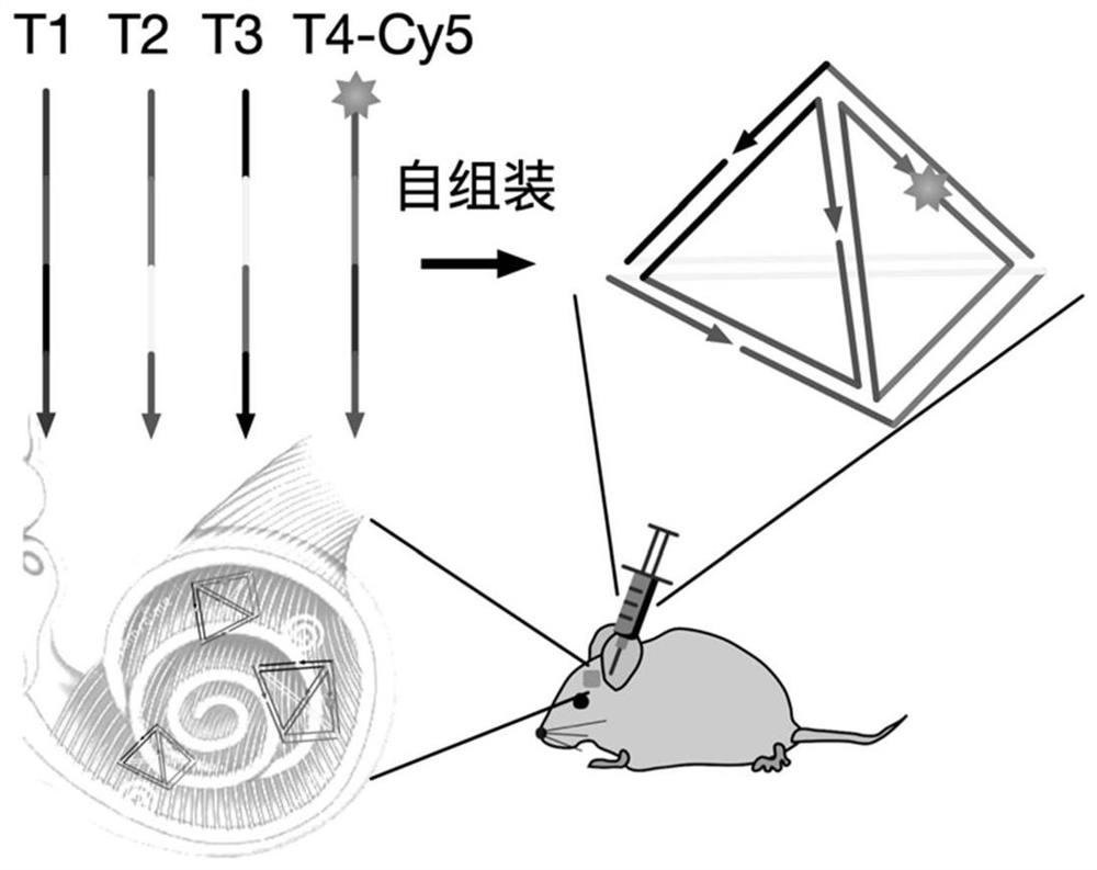
[0033] Example 1: Round window injection of neonatal mice
[0034] Neonatal mice from P2 to P3 were anesthetized with hypothermia for 2-3 minutes until they lost consciousness, and then moved to an ice pad for subsequent operations. The operation time was limited to 5-10min, and the operation was performed on the left ear of each animal. After anesthesia, a retroauricular incision was made to expose the bulla and the cochlea was observed. According to the relative position of temporal bone and facial nerve, the round window is exposed. Special care should be taken during the operation to avoid damage to the facial nerve. Using a glass microtube (25 μm) controlled by an ultramicropump, the DNA tetrahedral nanostructure solution was injected through the round window. The injection volume was controlled at 1.5-2 μL / cochlea. The negative control was injected with the same amount of Cy5 fluorophore solution. After injection, the skin incision is closed using veterinary tissue ...
Embodiment 2
[0035] Example 2: Cochlear tissue preparation and confocal imaging
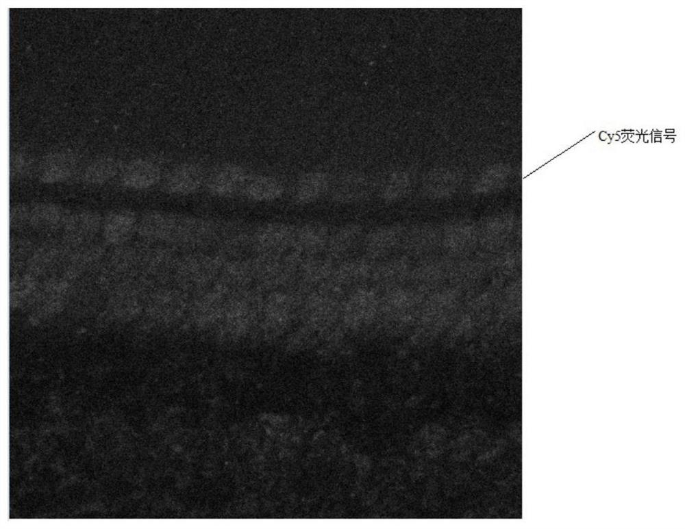
[0036]Mice were killed by cervical dislocation after anesthesia, and the inner ear was carefully removed after decapitation, and transferred to a fixative solution containing 4% paraformaldehyde. The excess tissue of the inner ear was removed under a stereomicroscope, and the round window, oval window, and cochlear top bone wall were opened with a needle. Suction and blow gently into the middle cochlea. Fix at room temperature for 2 hours, rinse twice with PBS solution to remove residual fixative solution, transfer to 10% EDTA-2Na decalcification solution for 72 hours at 4°C, and change the solution every day. After the inner ear of the mouse was fully decalcified, it was transferred to a glass dish filled with PBS buffer, and the decalcified volute was carefully removed under a stereomicroscope, and the basement membrane was gradually separated from the apical gyrus to the apical gyrus, and the surrounding i...
Embodiment 3
[0045] Example 3: Preparation of DNA tetrahedron nanostructure The framework nucleic acid tetrahedron provided by the present invention is synthesized from 4 DNA single strands, which were synthesized by Shanghai Shenggong Company and named T1, T2, T3, T4-Cy5 respectively. Among them, T1, T2, and T3 are ordinary nucleotide sequences without any chemical group modification. The 5' port of T4-Cy5 is modified with a Cy5 fluorophore to allow visualization of the tetrahedral position using confocal fluorescence microscopy. The numbers of DNA single strands are T1, T2, T3, T4-Cy5 respectively, and the specific sequences are shown in Table 1:
[0046] Table 1: DNA tetrahedron sequence design
[0047]
[0048] Concrete preparation method is:
[0049] (1) Dissolve and measure the concentration of DNA single strands ordered in dry powder state with numbers T1, T2, T3, T4-Cy5.
[0050] First, centrifuge the dry-fractionated DNA molecules numbered T1, the specific operation is to ce...
PUM
 Abstract
Description
Claims
Application Information
 - R&D
- Intellectual Property
- Life Sciences
- Materials
- Tech Scout
- Unparalleled Data Quality
- Higher Quality Content
- 60% Fewer Hallucinations
Browse by: Latest US Patents, China's latest patents, Technical Efficacy Thesaurus, Application Domain, Technology Topic, Popular Technical Reports.
© 2025 PatSnap. All rights reserved.Legal|Privacy policy|Modern Slavery Act Transparency Statement|Sitemap|About US| Contact US: help@patsnap.com


